. 2017 May 16;8(3):e00382-17.
doi: 10.1128/mBio.00382-17.

Conformational Flexibility in the Immunoglobulin-Like Domain of the Hepatitis C Virus Glycoprotein E2

Affiliations

Conformational Flexibility in the Immunoglobulin-Like Domain of the Hepatitis C Virus Glycoprotein E2

Ieva Vasiliauskaite et al. mBio. .

Abstract

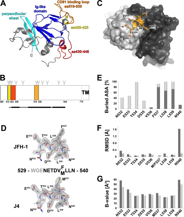
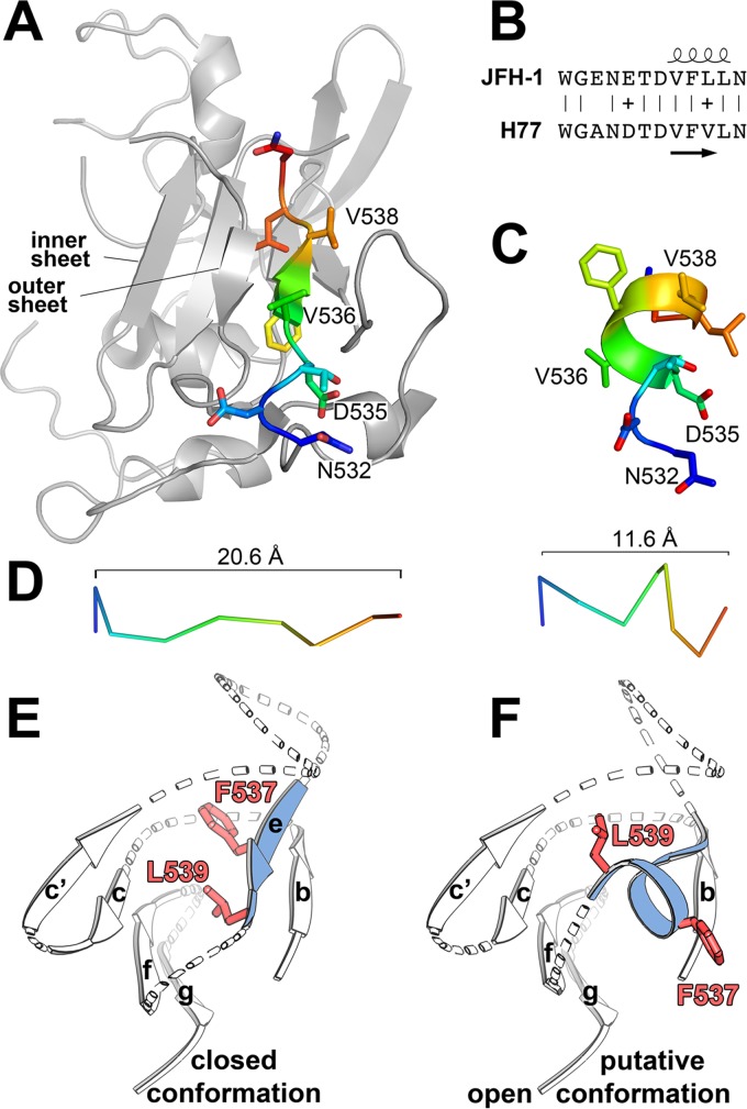
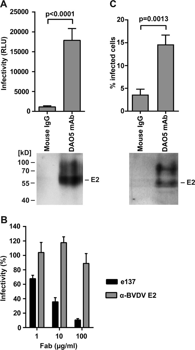
The hepatitis C virus (HCV) glycoprotein E2 is the major target of neutralizing antibodies and is therefore highly relevant for vaccine design. Its structure features a central immunoglobulin (Ig)-like β-sandwich that contributes to the binding site for the cellular receptor CD81. We show that a synthetic peptide corresponding to a β-strand of this Ig-like domain forms an α-helix in complex with the anti-E2 antibody DAO5, demonstrating an inside-out flip of hydrophobic residues and a secondary structure change in the composite CD81 binding site. A detailed interaction analysis of DAO5 and cross-competing neutralizing antibodies with soluble E2 revealed that the Ig-like domain is trapped by different antibodies in at least two distinct conformations. DAO5 specifically captures retrovirus particles bearing HCV glycoproteins (HCVpp) and infectious cell culture-derived HCV particles (HCVcc). Infection of cells by DAO5-captured HCVpp can be blocked by a cross-competing neutralizing antibody, indicating that a single virus particle simultaneously displays E2 molecules in more than one conformation on its surface. Such conformational plasticity of the HCV E2 receptor binding site has important implications for immunogen design.IMPORTANCE Recent advances in the treatment of hepatitis C virus (HCV) infection with direct-acting antiviral drugs have enabled the control of this major human pathogen. However, due to their high costs and limited accessibility in combination with the lack of awareness of the mostly asymptomatic infection, there is an unchanged urgent need for an effective vaccine. The viral glycoprotein E2 contains regions that are crucial for virus entry into the host cell, and antibodies that bind to these regions can neutralize infection. One of the major targets of neutralizing antibodies is the central immunoglobulin (Ig)-like domain within E2. We show here that this Ig-like domain is conformationally flexible at the surface of infectious HCV particles and pseudoparticles. Our study provides novel insights into the interactions of HCV E2 with the humoral immune system that should aid future vaccine development.

Keywords: CD81 binding site; Ig-like domain; conformational flexibility; glycoprotein E2; hepatitis C virus; monoclonal antibodies; vaccine design.


Figures

FIG 1
FIG 1
Crystal structure of DAO5 in complex with its peptide epitope. (A) Cartoon representation of cE2 (PDB 4WMF). The N and C termini are indicated; the Ig-like domain and the perpendicular sheet are highlighted in blue and cyan, respectively. The three segments contributing to the CD81 binding site are colored as described for panel B. (B) Linear diagram of the E2 glycoprotein. N-linked glycosylation sites are indicated above the diagram, and the three segments contributing to the composite CD81 binding site are shown as colored boxes and labeled with their amino acid residue numbers. A bar corresponding to the crystallized cE2 construct described in reference 10 is shown below the diagram, with the thinner line indicating disordered regions. (C) View of the DAO5 paratope in complex with the JFH-1 peptide (aa 529 to 540). The molecular surfaces of the light chain and heavy chain are colored light gray and dark gray, respectively, and the peptide is shown as a cartoon, displaying the side chains as sticks and colored according to atom type (orange, red, and blue for carbon, oxygen, and nitrogen, respectively). The α-helix formed by the C-terminal end of the peptide contacts the heavy chain CDR loops, while its N-terminal extended region (N532-T534) contacts the CDR loops of the light chain. (D) Electron density of the composite omit map contoured at 1σ around the peptide corresponding to HCV E2 strain JFH-1 (upper panel) or J4 (lower panel). The amino acid sequences of the peptides are displayed, with gray residues indicating disordered regions. (E) Percentages of accessible surface area (ASA) buried in the complex, calculated using PISA (51) and represented per residue as stacked columns for heavy (dark gray) and light (light gray) chains. (F) RMSD upon superposition of the two peptides, created using Chimera (52) and including all atoms (dark gray) or main chain atoms only (light gray) in the calculation and represented per residue. (G) Average temperature factors of the peptides plotted per residue for complexes with JFH-1 (light gray) and J4 (dark gray).
FIG 2
FIG 2
Conformation of the HCV E2 peptide aa 532 to 540. (A and C) Residues 532 to 540 of the extended conformation observed in the context of cE2 (PDB 4MWF) (A) and the helical conformation observed in the DAO5/peptide complex (C) are shown as cartoons, with the side chains shown as sticks and colored according to atom type (orange and red for oxygen and nitrogen, respectively). Carbon atoms are ramp colored from the N terminus (blue) to the C terminus (red) through green and yellow. cE2 is shown in gray. (B) Secondary structure elements taken from the crystal structures are shown above and below an amino acid alignment of the polypeptide segments from strains H77 and JFH-1. (D) Cα trace of the two backbone conformations colored as described for panel A. The extended conformation in the context of cE2 spans 20.6 Å (left panel), and the helical conformation spans 11.6 Å (right panel). (E and F) Schematic representation of the Ig-like domain within HCV E2 as observed in the E2 core fragment in the presence of a stabilizing Fab fragment (E) (PDB 4MWF) and in a putative open conformation (F) in which residues F537 and L539 (side chains are shown as red sticks) are solvent exposed, allowing for interaction with MAb DAO5, similar to the conformation of F537 observed in the second E2c structure (PDB 4NX3).
FIG 3
FIG 3
Cross-competition profile of DAO5. (A) Cross-competition and biochemical analysis of the sE2412-715–DAO5 complex. (Left) sE2412-715 and an sE2412-715–DAO5 scFv complex were affinity loaded onto a StrepTactin column, a CBH-4D Fab fragment was added, and the eluted complex was analyzed by SDS-PAGE under nonreducing conditions. (Right) CD81-LEL was incubated overnight at RT with the full-length HCV ectodomain (comprising residues 384 to 715; sE2) and the sE2-DAO5 Fab complex, followed by SEC and analysis of the peak fractions by SDS-PAGE under reducing conditions. DAO5 heavy and light chains form an apparent single band in the reducing gel due to an almost identical molecular mass of ~24 kDa. (B) A preformed sE2412-715–DAO5 Fab complex was incubated in the absence or presence of a Fab fragment targeting a non-overlapping (AP33) or overlapping (e137) region of cE2 and analyzed by SEC. After preincubation with AP33 (green), appearance of a peak at a higher molecular mass indicated ternary complex formation; after preincubation with e137 Fab (blue), the presence of peaks corresponding to the binary complex (at ~13 ml) and an isolated Fab fragment (at ~16 ml) showed that no ternary complex was formed. (C) Cross-competition profile of the sE2412-715–DAO5 Fab complex, obtained by SEC analysis as described for panel B, with a panel of Fab fragments derived from the indicated well-characterized anti-E2 MAbs. (D) sE2384-713 produced in human cells (eE2) was incubated with DAO5 Fab, and then the complex as well as the individual components were analyzed by SEC. The presence of a peak at a higher molecular mass indicated binary complex formation (12 ml, blue). A considerable protein fraction eluted in peaks corresponding to uncomplexed eE2 (green) and DAO5 Fab (red).
FIG 4
FIG 4
Evidence for more than one conformation of the CD81 binding loop. (A to C) sE2412-715 or sE2 was immobilized on a StrepTactin column and incubated first with a molar excess of DAO5 scFv and subsequently with e137 (A), AR3C (B), or A8 Fab (C). Eluted complexes were analyzed by SDS-PAGE. (D and E) Real-time SPR analysis of Fab binding to immobilized sE2 recorded the binding response (in resonance units [RU]) as a function of time. Fab fragments of e137 (D) and DAO5 (E) were injected over a surface with immobilized HCV sE2. After reaching equilibrium between association and dissociation, a second Fab fragment (either the same Fab again or the alternative one) or a buffer control was injected.
FIG 5
FIG 5
More than one conformation of the Ig-like domain was observed on both HCVpp and HCVcc infectious particles. (A) Concentrated HCVpp were immunocaptured using Immuno tubes. After removing unbound material, the captured particles were analyzed by SDS-PAGE, followed by immunoblotting to detect E2 (lower panel). The presence of captured infectious HCVpp was determined by overnight incubation with Huh7 cells in suspension. The cells were then transferred into tissue culture dishes, and infectivity levels were determined 48 h later by measuring luciferase activity, shown as relative light units (RLU) (upper panel). (B) DAO5-captured particles were incubated with Huh7 cells as described for panel A in the presence of e137 Fab or control anti-bovine viral diarrhea virus (BVDV) E2 Fab at the indicated concentrations. Infectivity levels were determined as described above and are shown as the percent infectivity in the absence of Fab. (C) Concentrated HCVcc were immunocaptured as described for panel A. After removing unbound material, captured particles were analyzed by SDS-PAGE followed by immunoblotting to detect E2 (lower panel). The presence of captured infectious HCVcc was determined by overnight incubation with Huh7 cells in suspension. The cells were then transferred into tissue culture dishes, and the number of infected cells was determined after 72 h by flow cytometry after labeling with anti-NS5A MAb 9E10 (upper panel). Experiments were performed at least in triplicate, and results shown are means ± standard deviations. The P values (panel A and C) were calculated using an unpaired t test.

References

    1. Shepard CW, Finelli L, Alter MJ. 2005. Global epidemiology of hepatitis C virus infection. Lancet Infect Dis 5:558–567. doi:10.1016/S1473-3099(05)70216-4. - DOI - PubMed
    1. Schinazi R, Halfon P, Marcellin P, Asselah T. 2014. HCV direct-acting antiviral agents: the best interferon-free combinations. Liver Int 34(Suppl 1):69–78. doi:10.1111/liv.12423. - DOI - PMC - PubMed
    1. Ball JK, Tarr AW, McKeating JA. 2014. The past, present and future of neutralizing antibodies for hepatitis C virus. Antiviral Res 105:100–111. doi:10.1016/j.antiviral.2014.02.013. - DOI - PMC - PubMed
    1. Pileri P, Uematsu Y, Campagnoli S, Galli G, Falugi F, Petracca R, Weiner AJ, Houghton M, Rosa D, Grandi G, Abrignani S. 1998. Binding of hepatitis C virus to CD81. Science 282:938–941. doi:10.1126/science.282.5390.938. - DOI - PubMed
    1. Scarselli E, Ansuini H, Cerino R, Roccasecca RM, Acali S, Filocamo G, Traboni C, Nicosia A, Cortese R, Vitelli A. 2002. The human scavenger receptor class B type I is a novel candidate receptor for the hepatitis C virus. EMBO J 21:5017–5025. doi:10.1093/emboj/cdf529. - DOI - PMC - PubMed

Publication types

MeSH terms

LinkOut - more resources