. 2017 May 16;8(3):e00646-17.
doi: 10.1128/mBio.00646-17.

Glutathionylation of Yersinia pestis LcrV and Its Effects on Plague Pathogenesis

Affiliations

Glutathionylation of Yersinia pestis LcrV and Its Effects on Plague Pathogenesis

Anthony Mitchell et al. mBio. .

Abstract

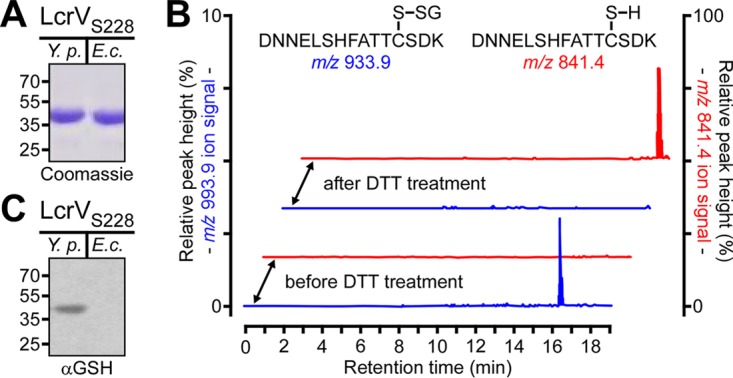
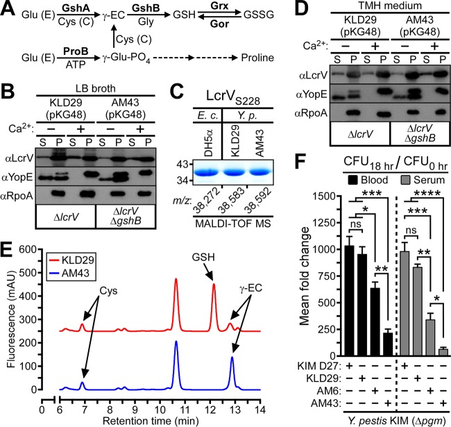
Glutathionylation, the formation of reversible mixed disulfides between glutathione and protein cysteine residues, is a posttranslational modification previously observed for intracellular proteins of bacteria. Here we show that Yersinia pestis LcrV, a secreted protein capping the type III secretion machine, is glutathionylated at Cys273 and that this modification promotes association with host ribosomal protein S3 (RPS3), moderates Y. pestis type III effector transport and killing of macrophages, and enhances bubonic plague pathogenesis in mice and rats. Secreted LcrV was purified and analyzed by mass spectrometry to reveal glutathionylation, a modification that is abolished by the codon substitution Cys273Ala in lcrV Moreover, the lcrVC273A mutation enhanced the survival of animals in models of bubonic plague. Investigating the molecular mechanism responsible for these virulence attributes, we identified macrophage RPS3 as a ligand of LcrV, an association that is perturbed by the Cys273Ala substitution. Furthermore, macrophages infected by the lcrVC273A variant displayed accelerated apoptotic death and diminished proinflammatory cytokine release. Deletion of gshB, which encodes glutathione synthetase of Y. pestis, resulted in undetectable levels of intracellular glutathione, and we used a Y. pestis ΔgshB mutant to characterize the biochemical pathway of LcrV glutathionylation, establishing that LcrV is modified after its transport to the type III needle via disulfide bond formation with extracellular oxidized glutathione.IMPORTANCEYersinia pestis, the causative agent of plague, has killed large segments of the human population; however, the molecular bases for the extraordinary virulence attributes of this pathogen are not well understood. We show here that LcrV, the cap protein of bacterial type III secretion needles, is modified by host glutathione and that this modification contributes to the high virulence of Y. pestis in mouse and rat models for bubonic plague. These data suggest that Y. pestis exploits glutathione in host tissues to activate a virulence strategy, thereby accelerating plague pathogenesis.

Keywords: Yersinia pestis; glutathionylation; innate immunity; macrophage; plague; ribosomal protein S3 (RPS3).


Figures

FIG 1
FIG 1
LcrV secreted by Y. pestis is glutathionylated at Cys273. (A) Coomassie-stained SDS-PAGE of LcrVS228 purified via Strep-Tactin affinity chromatography from either the culture supernatant of Y. pestis KLD29(pKG48) (Y.p.) or cell lysates of E. coli DH5α(pKG48) (E.c.). (B) Reconstructed ion traces for the unmodified (m/z 841.4) [(M+2H)2+ ion of 1,680.71] and glutathione-modified (m/z 993.9) [(M+2H)2+ ion of 1,985.79] tryptic peptides from the Y. pestis LcrVS228 protein encompassing Cys273 before and after treatment with dithiothreitol (DTT). The chromatograms show the absence of the unmodified peptides and the presence of the glutathione-modified peptides before treatment and following DTT treatment, the presence of the unmodified peptides, and the absence of the glutathione-modified peptides. (C) LcrVS228 purified from Y. pestis culture supernatants or E. coli extracts (rLcrVS228) was analyzed by immunoblotting with glutathione-specific antiserum (αGSH).
FIG 2
FIG 2
The lcrVC273A mutation abolishes LcrV glutathionylation and accelerates Y. pestis-mediated macrophage death. (A) Y. pestis KIM D27 lcrV, KLD29 ΔlcrV, and AM6 lcrVC273A were induced for type III secretion by growth at 37°C in M9-Casamino Acids (M9-Ca) minimal medium lacking exogenous calcium ions. Proteins in the supernatant (S) and bacterial pellet (P) were separated by centrifugation and identified by immunoblotting with rabbit antibodies specific for type III secretion substrates (LcrV and YopE), the secreted F1 pilus subunit (F1), and, as a fractionation control, cytoplasmic RNA polymerase subunit A (RpoA). (B) LcrV secreted by the indicated Y. pestis strains was assayed for glutathionylation by subjecting the culture supernatant to GST-Sepharose affinity chromatography. The load (L) and eluate (E) fractions were analyzed by immunoblotting with anti-LcrV. (C and D) Type III effector (YopM-Bla)-mediated cleavage of CCF2-AM-stained human polymorphonuclear leukocytes infected with Y. pestis KIM D27(pYopM-Bla), KLD29(pYopM-Bla), or AM6(pYopM-Bla) was analyzed via flow cytometry for blue fluorescence (YopM-Bla cleavage of CCF2-AM). (C) Representative histograms show the blue fluorescence traces of Y. pestis-infected neutrophils; the percentage of YopM-Bla-injected neutrophils (blue cells) is indicated above the gating scheme that was used to measure blue fluorescence above background. (D) Percentage of quantification of YopM-Bla-injected human neutrophils. Data are means ± standard errors of the means (SEM) (n = 3). *, P < 0.05, by two-tailed unpaired Student’s t test. (E and F) The kinetics of host cell death were examined by infecting murine J774.A1 macrophages with Y. pestis KIM D27 or AM6 and enumerating propidium iodide (PI)-positive cells. (E) Macrophages, either left uninfected or infected with Y. pestis KIM D27 or AM6, were stained with membrane-permeant (Hoechst [blue]) and membrane-impermeant (PI [red]) dyes and analyzed by fluorescence microscopy to determine Y. pestis-mediated macrophage death (PI positive, red, and Hoechst positive, magenta). (F) The kinetics of cell death were examined by quantifying PI-positive macrophages at timed intervals following Y. pestis infection. Data are means ± SEM (n = 3). *, P < 0.0001, and ns, not significant, by two-tailed unpaired Student’s t test.
FIG 3
FIG 3
Glutathionylation of LcrV enhances bubonic plague pathogenesis. (A) Survival of cohorts of BALB/c mice (n = 20) infected via subcutaneous (s.c.) inoculation with 20 CFU of Y. pestis CO92 lcrV or Y. pestis TD1 lcrVC273A. WT versus lcrVC273A, P < 0.0001. (B) Survival of cohorts of Brown Norway rats (n = 15) infected via subcutaneous inoculation with 500 CFU of Y. pestis CO92 or TD1. WT versus lcrVC273A, P = 0.0194. Statistical analysis was performed using the Gehan-Breslow-Wilcoxon test. Data are representative of two independent experiments.
FIG 4
FIG 4
LcrV binds to macrophage RPS3 and modulates host inflammatory responses. (A) Y. pestis-mediated death of J774.A1 macrophages was assessed 3 h postinfection by propidium iodide staining. (B) Y. pestis KLD29 lcrVS228(pKG48)-infected J774.A1 macrophages were subjected to Strep-Tactin affinity chromatography. The crude lysate (L), lysate supernatant (S), lysate pellet (P), flowthrough (FT), wash (W), and eluate (E) fractions were collected and analyzed by silver- and Coomassie-stained SDS-PAGE; bottom-up proteomics was used to identify the indicated protein bands as LcrVS228 and macrophage ribosomal protein S3 (RPS3). (C and D) Immunoblotting of lysate (L) and eluate (E) fractions generated during Strep-Tactin affinity chromatography reveals variable association between LcrV variants and RPS3 in J774.A1 macrophages infected with Y. pestis KLD29 ΔlcrV expressing wild-type lcrV (pNM77), lcrVS228 (pKG48), or lcrVS228 C273A (pAM128). (C) Representative immunoblot analysis of LcrV and RPS3 purified from Y. pestis-infected macrophages; actin levels were used as a loading control. (D) Densitometric quantification of the interaction between LcrVS228 and RPS3. Immunoreactive signals of Strep-Tactin-purified LcrV and RPS3 were normalized to the corresponding actin band; the ratio of eluate to lysate (E to L) is presented as a percent average. (E and F) Supernatants from J774.A1 macrophages, either left uninfected or infected with the indicated Y. pestis strains, were assayed by ELISA for (E) IL-1β or (F) IL-18. All data are means ± SEM (n = 3). **, P < 0.01; ***, P < 0.001; ****, P < 0.0001; ns, not significant.
FIG 5
FIG 5
Extracellular glutathione modifies secreted LcrV and promotes Y. pestis survival in blood. (A) Pathways of glutathione biosynthesis in E. coli. Glu (E), glutamic acid; Cys (C), cysteine; GshA, γ-glutamylcysteine synthetase; GshB, glutathione synthetase; ProB, γ-glutamyl kinase; γ-EC, γ-glutamylcysteine; GSH, glutathione; GSSG, glutathione disulfide; Gor, glutathione reductase; Grx, glutaredoxin; γ-glutamyl-PO4, γ-glutamyl phosphate. (B) Y. pestis KLD29 ΔlcrV lcrVS228(pKG48) and AM43 ΔlcrV ΔgshB lcrVS228(pKG48) were grown at 37°C in LB broth, in either the presence or absence of calcium ions (Ca2+), and assayed for type III secretion by immunoblotting; proteins secreted into the supernatant (S) were separated from intact bacteria (P) by centrifugation. (C) Y. pestis and E. coli strains expressing LcrVS228 (pKG48) were propagated in LB broth. LcrV was purified from Y. pestis supernatants (LcrVS228) or E. coli extracts (rLcrVS228), visualized by Coomassie-stained SDS-PAGE, and analyzed by MALDI-TOF MS to reveal the m/z of each LcrVS228 purification sample. (D) Y. pestis strains were analyzed for calcium-regulated type III secretion in TMH medium. (E) Partial HPLC chromatograms of LMW thiol-mBBr derivatives extracted from Y. pestis cultures grown to the stationary phase in TMH medium. Peaks corresponding to Cys, GSH, and γ-EC were assigned on the basis of retention time by comparison to a chromatogram of thiol standards. (F) Approximately 105 CFU of Y. pestis KIM D27 lcrV, KLD29 ΔlcrV, AM6 lcrVC273A, or AM43 ΔlcrV ΔgshB was inoculated into 4 ml of defibrinated sheep blood or heat-inactivated sheep serum. Culture aliquots were removed before and after 18 h of growth at 37°C and plated on LB agar to enumerate bacterial load. Y. pestis growth was calculated as the mean fold increase in bacteria at the time of inoculation (CFU0 h) to bacteria recovered after the 37°C incubation (CFU18 h). Data are means ± SEM (n = 3). *, P < 0.05, **, P < 0.01, ***, P < 0.001, and ****, P < 0.0001, and ns, not significant, by one-way ANOVA with Tukey’s multiple-comparison posttest.

Similar articles

Cited by

References

    1. Masip L, Veeravalli K, Georgiou G. 2006. The many faces of glutathione in bacteria. Antioxid Redox Signal 8:753–762. doi:10.1089/ars.2006.8.753. - DOI - PubMed
    1. Loi VV, Rossius M, Antelmann H. 2015. Redox regulation by reversible protein S-thiolation in bacteria. Front Microbiol 6:187. doi:10.3389/fmicb.2015.00187. - DOI - PMC - PubMed
    1. Dalle-Donne I, Rossi R, Colombo G, Giustarini D, Milzani A. 2009. Protein S-glutathionylation: a regulatory device from bacteria to humans. Trends Biochem Sci 34:85–96. doi:10.1016/j.tibs.2008.11.002. - DOI - PubMed
    1. Reniere ML, Whiteley AT, Hamilton KL, John SM, Lauer P, Brennan RG, Portnoy DA. 2015. Glutathione activates virulence gene expression of an intracellular pathogen. Nature 517:170–173. doi:10.1038/nature14029. - DOI - PMC - PubMed
    1. Potter AJ, Trappetti C, Paton JC. 2012. Streptococcus pneumoniae uses glutathione to defend against oxidative stress and metal ion toxicity. J Bacteriol 194:6248–6254. doi:10.1128/JB.01393-12. - DOI - PMC - PubMed

MeSH terms

LinkOut - more resources