

Anaemia and respiratory failure in a child: can it be idiopathic pulmonary haemosiderosis?
- PMID: 28512100
- PMCID: PMC5612269
- DOI: 10.1136/bcr-2017-219431
Anaemia and respiratory failure in a child: can it be idiopathic pulmonary haemosiderosis?
Abstract
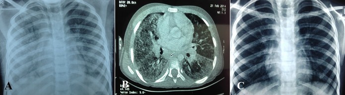
We present an 8-year-old male child admitted with cough and high-grade fever for 7 days and respiratory difficulty for 2 days. There was a history of blood transfusion at 2 years of age during a respiratory illness. The child was anaemic, tachycardic, tachypnoeic and hypoxic at presentation. Chest examination revealed equal air entry with fine crackles bilaterally. Blood reports were suggestive of anaemia (haemoglobin 6.5 g/dL), leucocytosis and high C reactive protein levels. Chest radiograph revealed bilateral air space opacities involving diffuse lung fields, right more than left. Relevant microbiological workup was negative. Based on the clinical scenario and investigations, a provisional diagnosis of pulmonary haemosiderosis was kept. The patient was started on intravenous pulse methylprednisolone. Fibre-optic bronchoscopy was done following recovery from the acute event. Bronchoalveolar lavage demonstrated a significant number of haemosiderin-laden macrophages confirming pulmonary haemosiderosis.
Keywords: Paediatrics; Respiratory medicine.
© BMJ Publishing Group Ltd (unless otherwise stated in the text of the article) 2017. All rights reserved. No commercial use is permitted unless otherwise expressly granted.
Conflict of interest statement
Competing interests: None declared.
Figures

References
-
- Sheikh SI, McCoy KS. Idiopathic pulmonary hemosiderosis : Taussig LM, Landau LI, Pediatric Respiratory Medicine. 2nd Edition Philadelphia: Mosby Elsevier, 2008.
-
- Kabra SK, Bhargava S, Lodha R, et al. Idiopathic pulmonary hemosiderosis: clinical profile and follow up of 26 children. Indian Pediatr 2007;44:333–8. - PubMed
-
- Vece TJ, De Guzman MM, Langston C, et al. Diffuse alveolar hemorrhage in children : Wilmott RW, Boat TF, Kendig and Chernick's disorders of the respiratory tract in children. 8th Edition Philadelphia: Elsevier Saunders, 2012.
Publication types
MeSH terms
Substances
LinkOut - more resources
Full Text Sources
Other Literature Sources
Medical
Research Materials