Comparative Study
. 2017 Jul 7;121(2):106-112.
doi: 10.1161/CIRCRESAHA.117.311212. Epub 2017 May 16.

Divergent Requirements for EZH1 in Heart Development Versus Regeneration

Affiliations
Comparative Study

Divergent Requirements for EZH1 in Heart Development Versus Regeneration

Shanshan Ai et al. Circ Res. .

Abstract

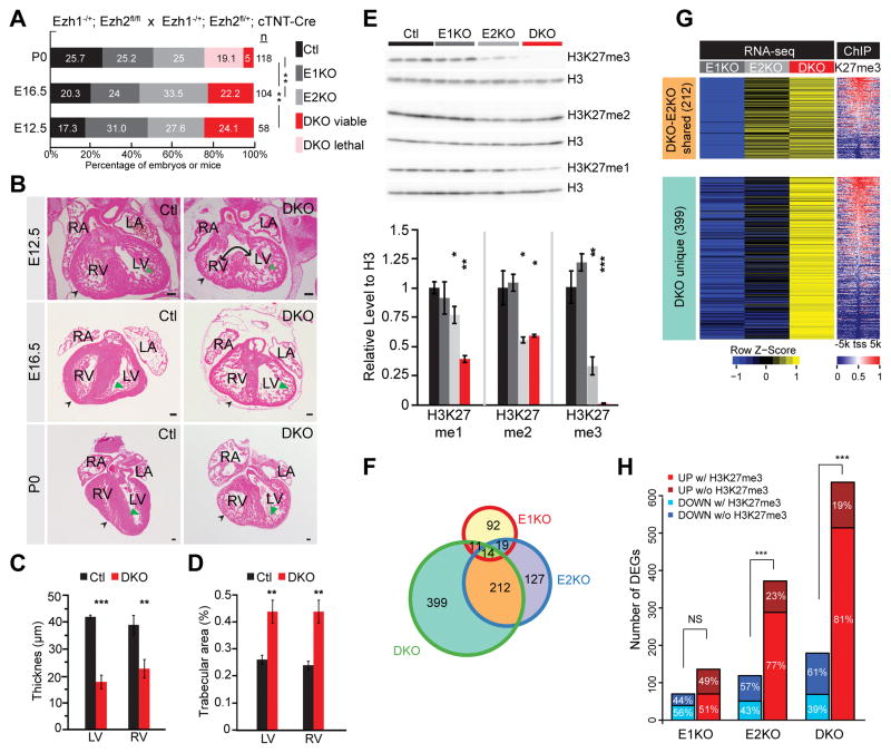
Rationale: Polycomb repressive complex 2 is a major epigenetic repressor that deposits methylation on histone H3 on lysine 27 (H3K27me) and controls differentiation and function of many cells, including cardiac myocytes. EZH1 and EZH2 are 2 alternative catalytic subunits with partial functional redundancy. The relative roles of EZH1 and EZH2 in heart development and regeneration are unknown.

Objective: We compared the roles of EZH1 versus EZH2 in heart development and neonatal heart regeneration.

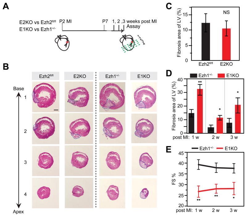
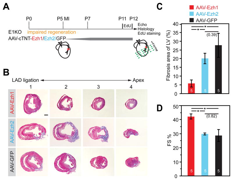
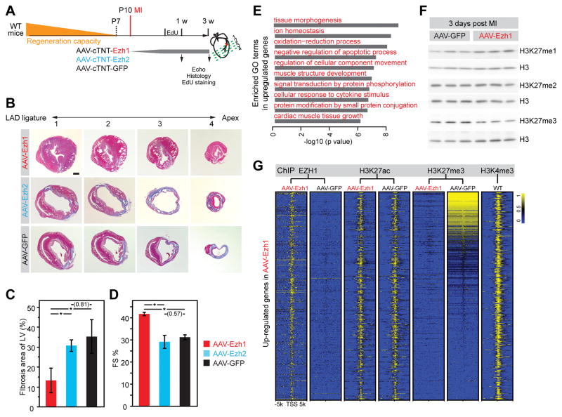
Methods and results: Heart development was normal in Ezh1-/- (Ezh1 knockout) and Ezh2f/f::cTNT-Cre (Ezh2 knockout) embryos. Ablation of both genes in Ezh1-/-::Ezh2f/f::cTNT-Cre embryos caused lethal heart malformations, including hypertrabeculation, compact myocardial hypoplasia, and ventricular septal defect. Epigenome and transcriptome profiling showed that derepressed genes were upregulated in a manner consistent with total EZH dose. In neonatal heart regeneration, Ezh1 was required, but Ezh2 was dispensable. This finding was further supported by rescue experiments: cardiac myocyte-restricted re-expression of EZH1 but not EZH2 restored neonatal heart regeneration in Ezh1 knockout. In myocardial infarction performed outside of the neonatal regenerative window, EZH1 but not EZH2 likewise improved heart function and stimulated cardiac myocyte proliferation. Mechanistically, EZH1 occupied and activated genes related to cardiac growth.

Conclusions: Our work unravels divergent mechanisms of EZH1 in heart development and regeneration, which will empower efforts to overcome epigenetic barriers to heart regeneration.

Keywords: lysine; methylation; myocardium; polycomb repressive complex 2; regeneration.


Conflict of interest statement

DISCLOSURES

The authors have no conflicts of interest to disclose.

Figures

Figure 1
Figure 1. Derangement of heart development and transcriptional regulation in cardiac Ezh1 and Ezh2 double knockout
A. Survival analysis of mutants at indicated developmental stages. A majority of DKO died perinatally. Numbers next to bars indicating sample size. P, postnatal. E, embryonic. Pearson’s Chi-squared test was used for P-value calculation. B. Cardiac abnormalities of E12.5, E16.5 and P0 embryos. Representative HE stained transverse sections revealing thinning of compact myocardium (black arrowheads), excessive myocardial trabeculation (green arrowheads), and ventricular septal defect (double-headed arrow). Scale bar = 100 μm.C and D. Quantification showing decreased compact myocardial thickness (C) and increased myocardial trabecular area (D). E. Western blotting and quantification showing downregulation of bulk H3K27me1/2/3 in DKO. Heart apex of embryos at E16.5 was used for protein extraction. F. Venn diagram showing differentially expressed genes in E1KO, E2KO and DKO compared to Ctl (Ezh1−/+ or Ezh2fl/+; cTNT-Cre). Differentially expressed genes identified by Cuffdiff (P<0.05 and log2(fold change)>0.5 or <−0.5). G. Heat map showing RNA-seq and H3K27me3 at ± 5 kb of TSS of genes in F. Genes are ordered by decreasing promoter H3K27me3 signal. H. Bar graph showing association of differentially expressed genes with H3K27me3 enrichment at ± 5 kb of TSS. RV, right ventricle; LV: left ventricle; RA, right atrium; LA, left atrium; CM, cardiac myocyte. *, P<0.05; **, P<0.01; ***, P<0.001 compared to control.
Figure 2
Figure 2. Ezh1 was required for neonatal heart regeneration after myocardial infarction
A. Experimental design and timeline. B–D. Representative Masson’s trichrome-stained cross-sections from E2KO, Ezh2fl/fl, Ezh1+/−, and E1KO hearts one week after MI (B) and quantification of fibrotic area of LV (C–D). E. Quantification of heart function by echocardiography. FS%, Fractional shortening. *, P<0.05; **P<0.01; n=7 per group. Scale bar = 500 μm.
Figure 3
Figure 3. Re-expression of Ezh1 but not Ezh2 restored cardiac function and regeneration in E1KO hearts
A. Schematic of rescue experimental design. AAV-cTNT-Ezh1/Ezh2/GFP were injected 24 hours subcutaneously before MI, and EdU was injected 24 hours before harvest. B–C. Representative Masson’s trichrome staining of cross-sections from E1KO mice treated with indicated AAV (B) and quantification of the percentage of the LV occupied by fibrotic scar (C). D. Echocardiographic measurement of heart function. FS%, fractional shortening. Scale bar = 500 μm; *, P<0.05. Numbers in parentheses indicate non-significant p-values. Numbers in bars indicate group size.
Figure 4
Figure 4. Over-expression of Ezh1 but not Ezh2 promoted heart regeneration by upregulating cardiac muscle growth genes
A. Schematic of rescue experimental design. AAV-cTNT-Ezh1, -Ezh2, or -GFP were injected subcutaneously 3 days before MI at P10, and EdU was injected 24 hours before harvest. Separate cohorts were analyzed at 1 week or 3 weeks post MI. B–C. Masson’s trichrome staining of cross-sections from hearts treated with AAV expressing indicated transgenes. B, representative sections. C, quantitative analysis. D. Echocardiographic measurement of heart function at 3 weeks post MI. FS%, fractional shortening. E. GO terms in which genes up-regulated in AAV-Ezh1-treated post MI hearts were enriched. F. Western blotting showing H3K27me1/2/3 bulk levels in hearts of mice injected with AAV-GFP or -Ezh1. G. Heat map showing H3K27me3, H3K27ac, EZH1 and H3K4me3 ChIP-seq signals at ± 5 kb of TSS of upregulated genes in AAV-Ezh1 compared to AAV-GFP. Scale bar = 500 μm; *, P<0.05. There were 4–6 replicates per group. Numbers in parentheses indicate non-significant P-values.

Comment in

References

    1. Bergmann O, Bhardwaj RD, Bernard S, Zdunek S, Barnabe-Heider F, Walsh S, Zupicich J, Alkass K, Buchholz BA, Druid H, Jovinge S, Frisen J. Evidence for cardiac myocyte renewal in humans. Science. 2009;324:98–102. - PMC - PubMed
    1. Ahuja P, Sdek P, MacLellan WR. Cardiac myocyte cell cycle control in development, disease, and regeneration. Physiol Rev. 2007;87:521–44. - PMC - PubMed
    1. Porrello ER, Mahmoud AI, Simpson E, Hill JA, Richardson JA, Olson EN, Sadek HA. Transient regenerative potential of the neonatal mouse heart. Science. 2011;331:1078–80. - PMC - PubMed
    1. Galdos FX, Guo Y, Paige SL, VanDusen NJ, Wu SM, Pu WT. Cardiac Regeneration: Lessons From Development. Circ Res. 2017;120:941–59. - PMC - PubMed
    1. Lin Z, von Gise A, Zhou P, Gu F, Ma Q, Jiang J, Yau AL, Buck JN, Gouin KA, van Gorp PR, Zhou B, Chen J, Seidman JG, Wang DZ, Pu WT. Cardiac-specific YAP activation improves cardiac function and survival in an experimental murine MI model. Circ Res. 2014;115:354–63. - PMC - PubMed

Publication types

Substances