. 2017 Jul 13;130(2):167-175.
doi: 10.1182/blood-2016-12-757823. Epub 2017 May 16.

Hematopoietic origin of Langerhans cell histiocytosis and Erdheim-Chester disease in adults

Affiliations

Hematopoietic origin of Langerhans cell histiocytosis and Erdheim-Chester disease in adults

Paul Milne et al. Blood. .

Abstract

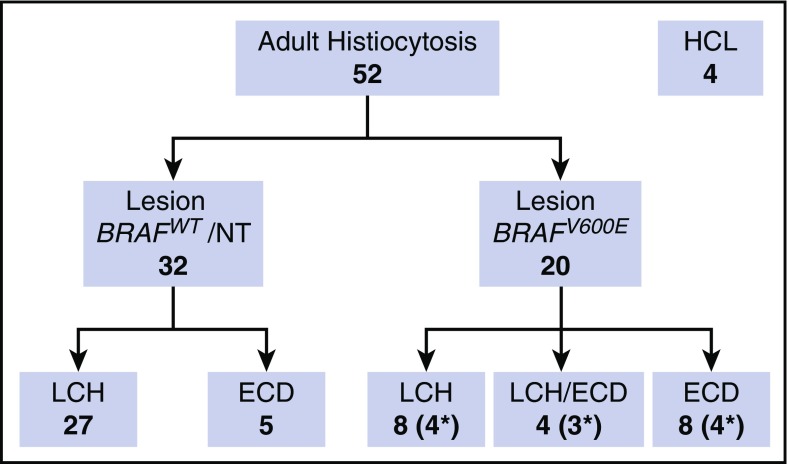
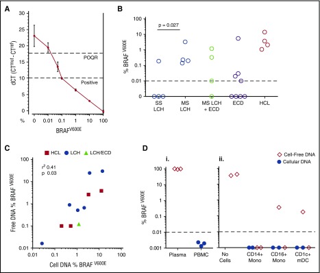
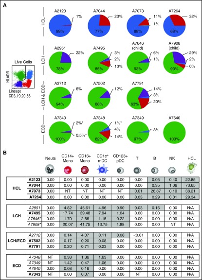
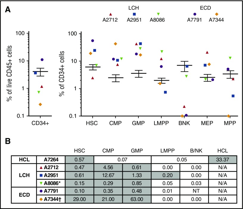
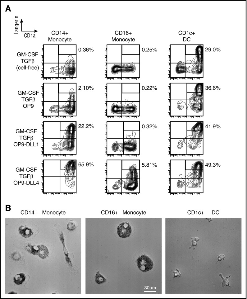
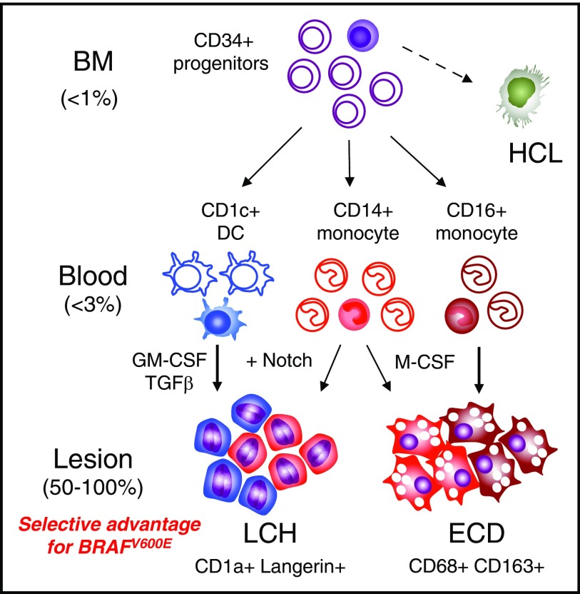
Langerhans cell histiocytosis (LCH) and Erdheim-Chester disease (ECD) are rare histiocytic disorders induced by somatic mutation of MAPK pathway genes. BRAFV600E mutation is the most common mutation in both conditions and also occurs in the hematopoietic neoplasm hairy cell leukemia (HCL). It is not known if adult LCH or ECD arises from hematopoietic stem cells (HSCs), nor which potential blood borne precursors lead to the formation of histiocytic lesions. In this study, BRAFV600E allele-specific polymerase chain reaction was used to map the neoplastic clone in 20 adults with LCH, ECD, and HCL. BRAFV600E was tracked to classical monocytes, nonclassical monocytes, and CD1c+ myeloid dendritic cells (DCs) in the blood, and mutations were observed in HSCs and myeloid progenitors in the bone marrow of 4 patients. The pattern of involvement of peripheral blood myeloid cells was indistinguishable between LCH and ECD, although the histiocytic disorders were distinct to HCL. As reported in children, detection of BRAFV600E in peripheral blood of adults was a marker of active multisystem LCH. The healthy counterparts of myeloid cells affected by BRAF mutation had a range of differentiation potentials depending on exogenous signals. CD1c+ DCs acquired high langerin and CD1a with granulocyte-macrophage colony-stimulating factor and transforming growth factor β alone, whereas CD14+ classical monocytes required additional notch ligation. Both classical and nonclassical monocytes, but not CD1c+ DCs, made foamy macrophages easily in vitro with macrophage colony-stimulating factor and human serum. These studies are consistent with a hematopoietic origin and >1 immediate cellular precursor in both LCH and ECD.


Figures

Figure 1.
Figure 1.
Patients. Summary of adult patients included in the study. Asterisks indicate the number of patients with BRAFV600E detectable in PBMCs. The BRAF wild-type (WT) ECD group includes 1 patient with RAS mutation. NT, not tested.
Figure 2.
Figure 2.
Detection of BRAFV600E alleles. (A) A standard curve constructed using DNA purified from a dilution series of BRAFV600E-positive melanoma cell line A375 into WT Epstein-Barr virus–transformed lymphoblastoid cell line. The quantitative limit of detection was 0.1%; the absolute limit of detection was 0.01% (positive outside of quantitative range [POQR]). (B) BRAFV600E allele frequency in bulk PBMCs in cases of lesion BRAFV600E + LCH, LCH/ECD, and ECD. Contingency of positive PBMCs upon MS-LCH tested by Fisher’s exact test. (C) Correlation between mutated allele burden in cell-free plasma and PBMC DNA in LCH, ECD, and HCL. (Di) Test of exogenous free DNA uptake by PBMCs. BRAF-mutated DNA (derived from melanoma cell line A375) was spiked into whole blood for 24 hours at room temperature. Plasma and PBMCs were isolated by density centrifugation. DNA was extracted from both fractions and subjected to allele-specific PCR. (Dii) Test of exogenous free DNA uptake by sorted cells incubated at 37°C for 24 hours in medium supplemented with 20% human serum from a patient with HCL. DNA from supernatants and cell pellets, as indicated, was subjected to allele-specific PCR. mDC, myeloid dendritic cell.
Figure 3.
Figure 3.
Allele-specific PCR of BRAFV600E in PBMCs. (A) The distribution of BRAFV600E in PBMCs sorted into quadrants as shown according to expression of HLA-DR and lineage. Pie charts summarize the distribution of mutant alleles in HCL, LCH, and ECD. The area of each quadrant in the pie is proportional to the total number of mutant alleles in that quadrant (ie, the percentage positivity of the quadrant multiplied by the number of cells in the quadrant). All patients had active disease at the time of sampling. Two children with LCH included for comparison are indicated. Asterisk indicates the threshold of detection for a negative result where cell numbers were limiting. (B) The distribution BRAFV600E among peripheral blood cells showing the percentage of mutated alleles detected. Gray shading indicates positive fractions. Asterisk indicates child with MS-LCH shown for comparison. All patients had active disease at the time of sampling. Italics indicate those who had received prior treatment. B, B cell; Mono, monocyte; Neuts, neutrophil; NK, natural killer; NT, not tested owing to lack of or insufficient material; pDC, plasmacytoid dendritic cell; T, T cell.
Figure 4.
Figure 4.
Mononuclear profile and BRAF allele frequency in CD34+ BM progenitors. (A) Relative proportions of progenitor fractions among CD34+ BM mononuclear cells compared with healthy controls (n = 21), expressed as percentage of total live cells. Error bars depict 95% confidence intervals of healthy controls. Populations: B/NK, B/NK cell progenitors; LMPP, lymphoid-primed multipotent progenitors; MEP, megakaryocyte–erythroid progenitor; MPP, multipotent progenitor. (B) The distribution of BRAFV600E among CD34+ BM progenitors. Gray shading indicates positive fractions. BM aspirate was obtained in parallel with the peripheral blood shown previously. *Child; NRASQ61R Sanger sequencing. NT, not tested owing to insufficient number of cells.
Figure 5.
Figure 5.
In vitro differentiation potential of monocytes and myeloid DCs. (A) Sorted CD14+ monocytes, CD16+ monocytes, and CD1c+ myeloid DCs from healthy controls cultured for 3 days in conditions as shown. LangerinhighCD1a+ gates depicted contain LCH-like cells with Birbeck granules. Notch ligands DLL1 And DLL4 were presented on transfected mouse OP9 cells. Representative data of 5 independent experiments. (B) Sorted CD14+ monocytes, CD16+ monocytes, and CD1c+ myeloid DCs from healthy controls cultured for 7 days with M-CSF and 5% human serum (HS) to reveal potential to differentiate into foamy macrophages. Representative phase contrast images of 5 independent experiments.
Figure 6.
Figure 6.
Potential precursor pathways in MS-LCH and ECD. Schema summarizing potential precursor pathways in MS-LCH and ECD based on the distribution of BRAFV600E alleles in peripheral blood and the differentiation potentials of healthy control cells in vitro. A minority of cells (filled) in the BM and blood contain BRAF mutation. The principal observation is that <1% clonal hematopoiesis gives rise to <3% mutated blood precursors that appear to be strongly selected for in peripheral tissue environments, resulting in lesional histiocyte mutation levels of up to 100%. Lesions potentially consist of >1 precursor as indicated by blue for CD1c+ DCs, red for classical monocytes, and brown for nonclassical monocytes. The depiction of lesion composition is conjectural and not based on experimental observation. Furthermore, additional contributions from other, more primitive myeloid progenitors cannot be excluded at present.

References

    1. Badalian-Very G, Vergilio JA, Degar BA, et al. . Recurrent BRAF mutations in Langerhans cell histiocytosis. Blood. 2010;116(11):1919-1923. - PMC - PubMed
    1. Sahm F, Capper D, Preusser M, et al. . BRAFV600E mutant protein is expressed in cells of variable maturation in Langerhans cell histiocytosis. Blood. 2012;120(12):e28-e34. - PubMed
    1. Haroche J, Charlotte F, Arnaud L, et al. . High prevalence of BRAF V600E mutations in Erdheim-Chester disease but not in other non-Langerhans cell histiocytoses. Blood. 2012;120(13):2700-2703. - PubMed
    1. Satoh T, Smith A, Sarde A, et al. . B-RAF mutant alleles associated with Langerhans cell histiocytosis, a granulomatous pediatric disease[published correction appears in PLoS One. 2012;7(6). doi:10.1371/annotation/74a67f4e-a536-4b3f-a350-9a4c1e6bebbd]. PLoS One. 2012;7(4):e33891. - PMC - PubMed
    1. Berres ML, Lim KP, Peters T, et al. . BRAF-V600E expression in precursor versus differentiated dendritic cells defines clinically distinct LCH risk groups[published correction appears in J. Exp. Med 2015;212(2):281. J Exp Med. 2014;211(4):669-683. - PMC - PubMed

Publication types

MeSH terms