Comparative Study
. 2017 Jun 6;474(12):2027-2038.
doi: 10.1042/BCJ20170097.

The mechanism of activation of IRAK1 and IRAK4 by interleukin-1 and Toll-like receptor agonists

Affiliations
Comparative Study

The mechanism of activation of IRAK1 and IRAK4 by interleukin-1 and Toll-like receptor agonists

Stefan Vollmer et al. Biochem J. .

Abstract

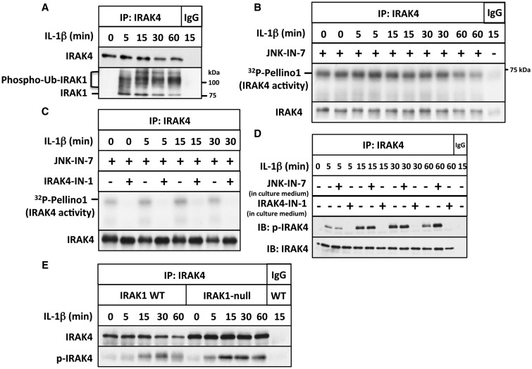
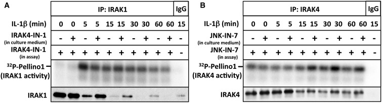
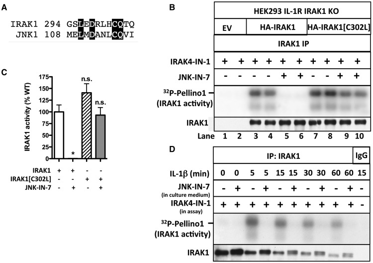
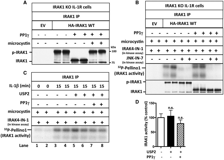
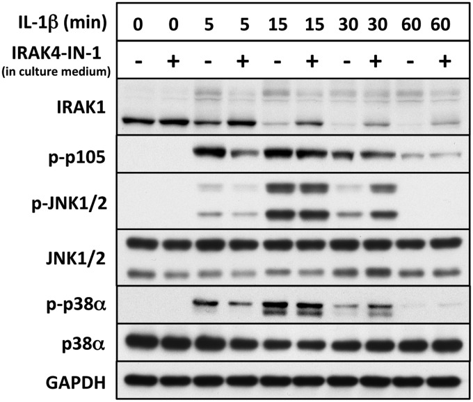
We have developed the first assays that measure the protein kinase activities of interleukin-1 receptor-associated kinase 1 (IRAK1) and IRAK4 reliably in human cell extracts, by employing Pellino1 as a substrate in conjunction with specific pharmacological inhibitors of IRAK1 and IRAK4. We exploited these assays to show that IRAK4 was constitutively active and that its intrinsic activity towards Pellino1 was not increased significantly by stimulation with interleukin-1 (IL-1) in IL-1R-expressing HEK293 cells, Pam3CSK4-stimulated human THP1 monocytes or primary human macrophages. Our results, in conjunction with those of other investigators, suggest that the IL-1-stimulated trans-autophosphorylation of IRAK4 is initiated by the myeloid differentiation primary response gene 88-induced dimerization of IRAK4 and is not caused by an increase in the intrinsic catalytic activity of IRAK4. In contrast with IRAK4, we found that IRAK1 was inactive in unstimulated cells and converted into an active protein kinase in response to IL-1 or Pam3CSK4 in human cells. Surprisingly, the IL-1-stimulated activation of IRAK1 was not affected by pharmacological inhibition of IRAK4 and not reversed by dephosphorylation and/or deubiquitylation, suggesting that IRAK1 catalytic activity is not triggered by a covalent modification but by an allosteric mechanism induced by its interaction with IRAK4.

Keywords: IRAK; MyD88; Pellino; Toll-like receptor; innate immunity.


Conflict of interest statement

The Authors declare that there are no competing interests associated with the manuscript.

Figures

Figure 1.
Figure 1.. IL-1 stimulates the autophosphorylation but not the activation of IRAK4.
(A) IL-1R cells were stimulated with IL-1β for the times indicated; IRAK4 immunoprecipitated from 0.5 mg of cell extract protein using anti-IRAK4 or control preimmune IgG. The presence of IRAK4 and that of IRAK1 were detected by immunoblotting, and phosphorylated and ubiquitylated forms of IRAK1 are denoted by phospho-Ub-IRAK1. (B) Same as in A, except that 1 mg of cell extract protein was used and the immunoprecipitates were assayed for IRAK4 activity using GST-Pellino1 and Mg[γ32P-ATP] as substrates in the presence (+) or absence (−) of the IRAK1 inhibitor JNK-IN-7 (1 µM). The figure shows an autoradiograph of the 32P-labelled Pellino1 formed during the assay. The radioactive band migrating more rapidly than GST-Pellino1 is a minor proteolytic fragment present in the preparation. The membranes were also immunoblotted for IRAK4. (C) Same as in B, except that IRAK4 was assayed in the presence of JNK-IN-7 (1 µM) and in the absence (−) or presence (+) of the IRAK4 inhibitor IRAK4-IN-1 (1 µM). (D) Same as in A, except that the cells were incubated for 1 h in the absence (−) or presence (+) of JNK-IN-7 (10 µM) or IRAK4-IN-1 (3 µM) prior to stimulation with IL-1β, and immunoblotting was carried out with antibodies that recognize all forms of IRAK4 (IRAK4) or antibodies that recognize IRAK4 phosphorylated at Thr345 and Ser346 (p-IRAK4). (E) Same as in D, except that IRAK4 was immunoprecipitated from both IL-1R cells and IRAK1-null IL-1R cells and no inhibitors were present in the cell culture medium.
Figure 2.
Figure 2.. IL-1 induces the activation of IRAK1.
(A) Same as in Figure 1B, except that IRAK1 was immunoprecipitated with anti-IRAK1 or control IgG and the IPs were assayed for IRAK1 activity using GST-Pellino1 and Mg[γ32P-ATP] as substrates in the presence (+) of IRAK4-IN-1 to inhibit co-immunoprecipitating IRAK4, and in the absence (−) or presence (+) of the IRAK1 inhibitor JNK-IN-7 (1 µM). (B) Same as in A, except that IRAK1 was assayed in the absence (−) or presence (+) of JNK-IN-7 (1 µM) or JNK-IN-8 (1 µM), and the membranes were additionally immunoblotted for all forms of IRAK1 — note that the unmodified form of IRAK1 disappears as it is gradually converted into more slowly migrating phosphorylated and ubiquitylated species after stimulation with IL-1β. (C) Same as in B, except that IRAK1 was immunoprecipitated from extracts of both IL-1R cells and IRAK1 KO IL-1R cells, and JNK-IN-7 was omitted from all assays. (D) Same as in B, except that IRAK4 was immunoprecipitated, and IRAK1 in the immunoprecipitates was assayed by including IRAK4-IN-1 in the assays.
Figure 3.
Figure 3.. The IL-1-stimulated activation of IRAK1 does not require the catalytic activity of IRAK4.
(A) IL-1R cells were incubated for 1 h without (−) or with (+) 3 µM IRAK4-IN-1 to inhibit cellular IRAK4 and stimulated with IL-1β for the times indicated. IRAK1 was immunoprecipitated from 1 mg of cell extract protein with anti-IRAK1 or control IgG and assayed with GST-Pellino1 and Mg[γ32P-ATP] as substrates in the presence (+) of 1 µM IRAK4-IN-1. Further details are given in Figure 2A. (B) Same as in A, except that the cell culture medium was incubated with 10 µM JNK-IN-7 to inactivate cellular IRAK1 activity, and IRAK4 was immunoprecipitated from the cell extracts instead of IRAK1, and co-immunoprecipitating IRAK4 was assayed by including JNK-IN-7 in the assay to inhibit IRAK1.
Figure 4.
Figure 4.. JNK-IN-7 is an irreversible inhibitor of IRAK1.
(A) The amino acid sequences of human IRAK1 (residues 294–305) and human JNK1 (residues 108–119) were aligned, and conserved residues are highlighted. Met108 of JNK1 is the ‘gatekeeper’ residue. (B) IRAK1 KO IL-1R cells were transfected with 5 µg or DNA of a control empty vector (EV) (lanes 1 and 2), HA-tagged wild-type IRAK1 (lanes 3–6) or HA-tagged IRAK1[C302L] (lanes 7–10). IRAK1 was immunoprecipitated from 0.5 mg of cell extract protein and assayed with GST-Pellino1 and Mg[γ32P-ATP] in the presence of IRAK4-IN-1 to inhibit co-immunoprecipitating IRAK4 and in the presence or absence of the IRAK1 inhibitor JNK-IN-7. (C) The autoradiogram from B and two other independent experiments were scanned, and the activity of IRAK1 and IRAK1[C302L] (IRAK1[C/L]) measured without or with 1.0 µM JNK-IN-7. The results are shown as a % of wild-type IRAK1 activity in the absence of JNK-IN-7. (D) IL-1R cells were incubated for 1 h without (−) or with (+) 10 µM JNK-IN-7 to inhibit cellular IRAK1 and then stimulated with IL-1β. IRAK1 was immunoprecipitated from 1 mg of cell extract protein and assayed with GST-Pellino1 and Mg[γ32P-ATP] as substrates in the presence (+) of 1 µM IRAK4-IN-1 to inhibit co-immunoprecipitating IRAK4.
Figure 5.
Figure 5.. IRAK1 activation does not require its phosphorylation or ubiquitylation.
(A) IRAK1 KO IL-1R cells were transfected with 5 µg of DNA of control empty vector (EV) or HA-IRAK1, and IRAK1 then immunoprecipitated from 0.5 mg of cell extract protein. The IPs were incubated with PP1γ (10 U) in the presence or absence of microcystin (10 µM) and denatured in SDS. Following SDS–PAGE and transfer to PVDF membranes, immunoblotting was performed with anti-IRAK1. (B) As in A, except that, after PP1γ treatment, the immunoprecipitates were washed, incubated for 1 h with 10 µM microcystin, and IRAK1 was assayed with GST-Pellino1 and Mg[γ32P-ATP] as substrates in the absence (−) or presence (+) of JNK-IN-7 (1 µM) and in the presence (+) of IRAK4-IN-1 (1 µM) to inactivate co-immunoprecipitating IRAK4. The presence of IRAK1 in the immunoprecipitates was also analyzed by immunoblotting. (C) IL-1R cells were stimulated with IL-1β and IRAK1 immunoprecipitated from 1 mg of cell extract protein and incubated with PP1γ (10 U) and USP2 (1.15 µg). The immunoprecipitates were washed, incubated for 1 h with 1 µM microcystin to inactivate any residual PP1γ, and IRAK1 assayed with GST-Pellino1 and Mg[γ32P-ATP] as substrates in the presence (+) of IRAK4-IN-1 (1 µM) to inactivate any co-immunoprecipitating IRAK4. (D) The autoradiogram from C and one other independent experiment were scanned and IRAK1 activity quantitated after incubation with or without USP2 and PP1γ. The results are presented as a % of that measured without USP2 and PP1γ treatment.
Figure 6.
Figure 6.. The inhibition of IRAK4 does not prevent the IL-1-stimulated activation of IKKβ and MAP kinases in IL-1R cells.
IL-1R cells were incubated for 1 h without (−) or with (+) 3 µM IRAK4-IN-1 to inactivate cellular IRAK4, and then stimulated with IL-1β. Cell extract (20 µg of protein) was subjected to SDS–PAGE and immunoblotting with antibodies recognizing all forms of IRAK1, JNK, p38α MAP kinase and GAPDH, and antibodies that recognize the phosphorylated (p) forms of p105/NF-κB1, JNK1/2 and p38α MAP kinase.

References

    1. Kawai T. and Akira S. (2010) The role of pattern-recognition receptors in innate immunity: update on Toll-like receptors. Nat. Immunol. 11, 373–384 doi:10.1038/ni.1863 - DOI - PubMed
    1. Lin S.-C., Lo Y.-C. and Wu H. (2010) Helical assembly in the MyD88–IRAK4–IRAK2 complex in TLR/IL-1R signalling. Nature 465, 885–890 doi:10.1038/nature09121 - DOI - PMC - PubMed
    1. Motshwene P.G., Moncrieffe M.C., Grossmann J.G., Kao C., Ayaluru M., Sandercock A.M. et al. (2009) An oligomeric signaling platform formed by the Toll-like receptor signal transducers MyD88 and IRAK-4. J. Biol. Chem. 284, 25404–25411 doi:10.1074/jbc.M109.022392 - DOI - PMC - PubMed
    1. Flannery S. and Bowie A.G. (2010) The interleukin-1 receptor-associated kinases: critical regulators of innate immune signalling. Biochem. Pharmacol. 80, 1981–1991 doi:10.1016/j.bcp.2010.06.020 - DOI - PubMed
    1. Pauls E., Nanda S.K., Smith H., Toth R., Arthur J.S.C. and Cohen P. (2013) Two phases of inflammatory mediator production defined by the study of IRAK2 and IRAK1 knock-in mice. J. Immunol. 191, 2717–2730 doi:10.4049/jimmunol.1203268 - DOI - PMC - PubMed

Publication types

MeSH terms