. 2017 Apr 15;31(8):830-844.
doi: 10.1101/gad.295741.116.

Regulation of DNA demethylation by the XPC DNA repair complex in somatic and pluripotent stem cells

Affiliations

Regulation of DNA demethylation by the XPC DNA repair complex in somatic and pluripotent stem cells

Jaclyn J Ho et al. Genes Dev. .

Abstract

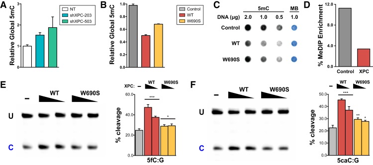
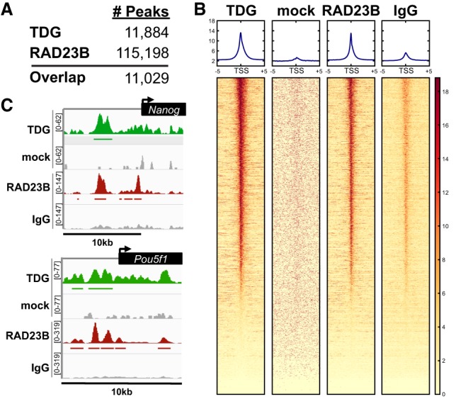
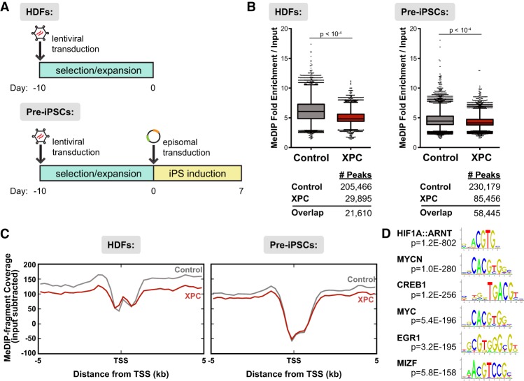
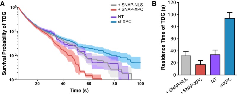
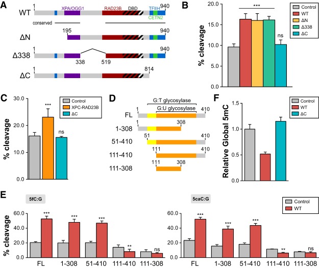
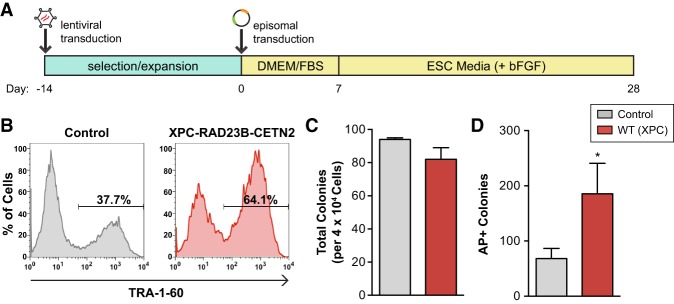
Faithful resetting of the epigenetic memory of a somatic cell to a pluripotent state during cellular reprogramming requires DNA methylation to silence somatic gene expression and dynamic DNA demethylation to activate pluripotency gene transcription. The removal of methylated cytosines requires the base excision repair enzyme TDG, but the mechanism by which TDG-dependent DNA demethylation occurs in a rapid and site-specific manner remains unclear. Here we show that the XPC DNA repair complex is a potent accelerator of global and locus-specific DNA demethylation in somatic and pluripotent stem cells. XPC cooperates with TDG genome-wide to stimulate the turnover of essential intermediates by overcoming slow TDG-abasic product dissociation during active DNA demethylation. We further establish that DNA demethylation induced by XPC expression in somatic cells overcomes an early epigenetic barrier in cellular reprogramming and facilitates the generation of more robust induced pluripotent stem cells, characterized by enhanced pluripotency-associated gene expression and self-renewal capacity. Taken together with our previous studies establishing the XPC complex as a transcriptional coactivator, our findings underscore two distinct but complementary mechanisms by which XPC influences gene regulation by coordinating efficient TDG-mediated DNA demethylation along with active transcription during somatic cell reprogramming.

Keywords: DNA methylation; epigenetics; pluripotency; reprogramming.


Figures

Figure 1.
Figure 1.
Global DNA methylation is inversely correlated with XPC expression independent of DNA repair activity. Relative global DNA methylation was assayed by 5mC-specific ELISA using genomic DNA from XPC knockdown H9 human ESCs (A) and HDFs overexpressing wild-type (WT) or DNA repair-deficient (W690S) human XPC (B). Relative global DNA methylation in HDFs was also assayed by 5mC dot blot (C) and MeDIP enrichment (D). Methylene blue (MB) staining was used to control for total DNA transferred to the membrane. (E,F) TDG cleavage activity of a 5′-labeled 37mer double-stranded oligonucleotide DNA (0.2 µM) in the presence or absence of decreasing amounts of wild-type or W690S mutant XPC (0.2–0.4 µM). The 37mer dsDNA contains either a 5fC:G (E) or 5caC:G (F) base pair as substrate for TDG. Uncleaved intact 37mer (U) and its cleaved product (C) were separated on a denaturing PAGE gel. Representative gels are shown. The intensity of the cleaved product indicates the efficiency of base excision by TDG and is calculated in the graphs as the percentage of total labeled substrate [=C/(U + C)]. Error bars represent the standard deviation. n = 3. (***) P < 0.001; (**) P < 0.01; (*) P < 0.05, calculated by two-way ANOVA.
Figure 2.
Figure 2.
TDG is enriched at enhancers and promoters genome-wide and colocalizes with the XPC complex subunit RAD23B. (A) The number of ChIP-seq peaks identified using MACS2 for each data set. Approximately 93% of biotin–TDG (Neri et al. 2015) ChIP-seq peaks overlap with RAD23B in mouse ESCs. (B) Heat map of TDG- and RAD23B-bound regions over a ±5-kb window around the TSSs of genes. Genes in each heat map are sorted according to descending levels of TDG ChIP-seq signal. (C) Integrative Genomics Viewer (IGV)-computed ChIP-seq tracks are plotted as (number of reads) × [1,000,000/(total read count)] for pluripotency genes Nanog and Pou5f1. Mock and normal IgG were used as specificity controls for the TDG and RAD23B ChIP, respectively.
Figure 3.
Figure 3.
Ectopic expression of XPC results in loss of DNA methylation genome-wide in both HDFs and pre-iPSCs. (A) Design of MeDIP-seq experiments. Reprogramming factors are cotransfected with a GFP-expressing plasmid to allow for sorting of transfected cells. (B) Up-regulation of XPC expression results in lower enrichment over background and fewer peaks called in MeDIP-seq analyses for both uninduced HDFs (left) and pre-iPSCs (right; 7 d post-induction). The number of peaks identified for each data set using MACS2 is shown below the graphs. (C) Distribution of MeDIP-seq reads by their distance ±5 kb from the TSSs of RefSeq genes, input subtracted. (D) Motif discovery of pre-iPSC MeDIP peaks enriched in the control but not the XPC gain-of-function data set.
Figure 4.
Figure 4.
XPC modulates TDG-binding dynamics to DNA in vivo. (A) Survival probability of an individual TDG-Halo molecule being bound as a function of time. Individual molecules were tracked using single-molecule imaging over consecutive frames and binned by their total length in cells cotransfected with SNAP-NLS or SNAP-XPC or transduced with lentiviruses containing a control NT shRNA or shRNA against XPC (shXPC). Bold lines represent the mean probability; shaded regions denote the 95% confidence interval. (B) Quantification of TDG DNA-bound residence time using a two-exponential decay model after correction for photobleaching using Halo-H2B (see Supplemental Fig. S7K). Error bars depict the standard deviation of the model fit.
Figure 5.
Figure 5.
XPC-mediated stimulation of TDG activity requires the C terminus of XPC and N terminus of TDG. (A) Schematic representation of human XPC (wild-type [WT]) highlighting the DNA-binding domain (DBD) and various protein–protein interaction domains. XPC truncations used in this study—ΔN (195–940 amino acids), Δ338 (del338–519 amino acids), and ΔC (1–814 amino acids)—are also shown. (B) TDG cleavage activity of a 5′-labeled T:G mismatch-containing oligonucleotide dsDNA in the presence of wild-type XPC or various XPC truncation complexes (ΔN, Δ338, and ΔC). (C) The CETN2 subunit is dispensable for stimulation of TDG-mediated base excision. TDG cleavage activity of a 5′-labeled T:G mismatch DNA substrate in the presence of the XPC–RAD23B heterodimer or a heterodimer containing ΔC XPC and RAD23B. (D) Schematic representation of full-length human TDG (FL) and truncations used in the in vitro glycosylase assay. The domains shown previously to be essential for G:U and G:T mismatch repair are indicated (Gallinari and Jiricny 1996). (E) Glycosylase activity of TDG truncations in the presence or absence of wild-type XPC was assayed using 5fC:G (left) and 5caC:G (right) substrates. The concentrations used were 0.2 µM DNA, 0.4 µM XPC complexes, and 50 nM TDG. (F) Relative global DNA methylation assayed by 5mC-specific ELISA using genomic DNA purified from HDFs overexpressing wild-type or ΔC human XPC. Error bars represent the standard deviation. n = 3. (***) P < 0.001; (**) P < 0.01; (n.s.) nonsignificant, calculated by two-way ANOVA.
Figure 6.
Figure 6.
XPC increases somatic reprogramming fidelity and iPSC self-renewal capacity. (A) Schematic diagram for human iPSC induction modified from methods in Okita et al. (2011). (B) Flow cytometry analysis of control and XPC complex (XPC–RAD23B–CETN2) gain-of-function cell populations 24 d post-induction for the late stage human iPSC marker TRA-1-60. (C) The average number of colonies obtained in the reprogramming experiment depicted in B. (D) iPSCs derived from control or XPC–RAD23B–CETN2-overexpressing HDFs were challenged with single-cell dissociation and allowed to recover for 3–4 d before staining with alkaline phosphatase (AP). The average number of AP+ colonies formed per 2.5 × 10⁴ single cells plated. Error bars depict the standard deviation. n = 3. (*) P < 0.05, calculated by two-tailed Student's t-test.

Similar articles

Cited by

References

    1. Afgan E, Baker D, van den Beek M, Blankenberg D, Bouvier D, Čech M, Chilton J, Clements D, Coraor N, Eberhard C, et al. 2016. The Galaxy platform for accessible, reproducible and collaborative biomedical analyses: 2016 update. Nucleic Acids Res 44: W3–W10. - PMC - PubMed
    1. Araki R, Hoki Y, Uda M, Nakamura M, Jincho Y, Tamura C, Sunayama M, Ando S, Sugiura M, Yoshida MA, et al. 2011. Crucial role of c-Myc in the generation of induced pluripotent stem cells. Stem Cells 29: 1362–1370. - PubMed
    1. Bailey TL, Boden M, Buske FA, Frith M, Grant CE, Clementi L, Ren J, Li WW, Noble WS. 2009. MEME suite: tools for motif discovery and searching. Nucleic Acids Res 37: W202–W208. - PMC - PubMed
    1. Bhutani N, Brady JJ, Damian M, Sacco A, Corbel SY, Blau HM. 2010. Reprogramming towards pluripotency requires AID-dependent DNA demethylation. Nature 463: 1042–1047. - PMC - PubMed
    1. Bird A. 2002. DNA methylation patterns and epigenetic memory. Genes Dev 16: 6–21. - PubMed

Publication types