. 2017 Jun 15;77(12):3151-3157.
doi: 10.1158/0008-5472.CAN-17-0035. Epub 2017 May 16.

Merkel Cell Polyomavirus Small T Antigen Initiates Merkel Cell Carcinoma-like Tumor Development in Mice

Affiliations

Merkel Cell Polyomavirus Small T Antigen Initiates Merkel Cell Carcinoma-like Tumor Development in Mice

Monique E Verhaegen et al. Cancer Res. .

Abstract

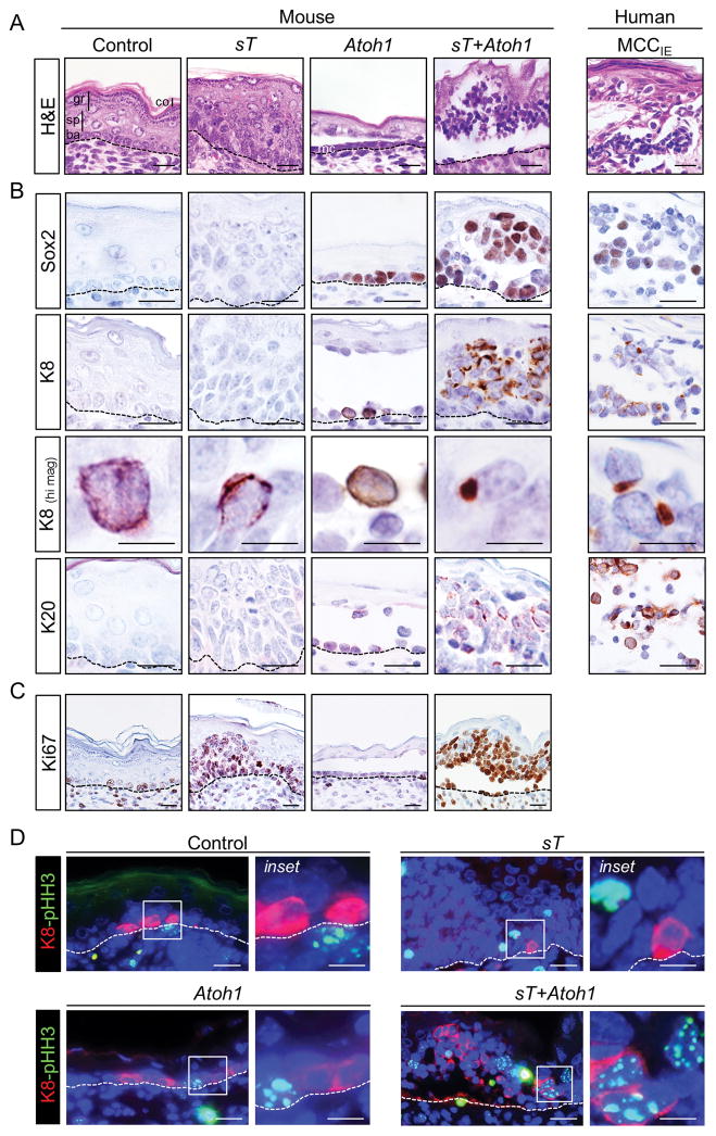
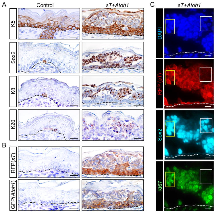
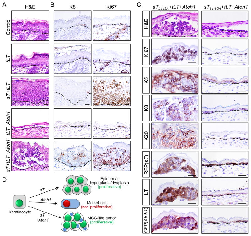
Merkel cell carcinoma (MCC) tumor cells express several markers detected in normal Merkel cells, a nonproliferative population of neuroendocrine cells that arise from epidermis. MCCs frequently contain Merkel cell polyomavirus (MCPyV) DNA and express viral transforming antigens, sT and tLT, but the role of these putative oncogenes in MCC development, and this tumor's cell of origin, are unknown. Using a panel of preterm transgenic mice, we show that epidermis-targeted coexpression of sT and the cell fate-determinant atonal bHLH transcription factor 1 (ATOH1) leads to development of widespread cellular aggregates, with histology and marker expression mimicking that of human intraepidermal MCC. The MCC-like tumor phenotype was dependent on the FBXW7-binding domain of sT, but not the sT-PP2A binding domain. Coexpression of MCPyV tLT did not appreciably alter the phenotype driven by either sT or sT combined with ATOH1. MCPyV sT, when coexpressed with ATOH1, is thus sufficient to initiate development of epidermis-derived MCC-like tumors in mice. Cancer Res; 77(12); 3151-7. ©2017 AACR.


Figures

Figure 1
Figure 1. MCPyV sT expression in ectopic Merkel cells initiates MCC-like tumor development in mice
A) Histology of epidermis from control and pre-term transgenic mouse embryos expressing MCPyV sT, Atoh1 or sT+Atoh1 driven by the Keratin 5 promoter compared to human intraepidermal MCC (MCCIE). Control epidermis contains a single layer of basal cells (ba) with several layers of differentiating spinous (sp), granular (gr) and cornified (co) cells. Expression of sT alone leads to epidermal hyperplasia and dysplasia, Atoh1 alone drives ectopic Merkel cells (mc) within a hypoplastic epidermis, whereas co-expression of sT and Atoh1 leads to MCC-like tumors. B) Immunohistochemical staining for Merkel cell and MCC markers Sox2, K8 and K20. Appearances of tumor-like aggregates of cells and of K8 in paranuclear dots is restricted to mice expressing sT+Atoh1. C) Ki67 immunostaining shows high proliferative activity in multiple layers of epidermis only in sT-expressing mice. D) Co-immunofluorescence for phospho-Histone H3 (pHH3) and K8 indicates mitotic activity of K8-positive cells largely restricted to MCC-like tumor cells in sT+Atoh1 mice. Scale bars = 25 μm; scale bars for K8 (hi mag) = 10 μm.
Figure 2
Figure 2. Sustained expression of Merkel cell markers in MCC-like mouse tumor cells with reduced levels of transgene expression
A) Immunohistochemical staining shows markedly reduced expression of K5 in suprabasal epidermis of sT+Atoh1 mice where MCC-like tumor cells express Merkel cell markers Sox2, K8 and K20. Isolated normal Merkel cells in control skin also express these markers (left panels). B) K5-driven expression of sT (RFP) and Atoh1 (GFP) transgenes is reduced in suprabasal regions of MCC-like tumors. C) Co-immunostaining shows robust Sox2 expression in regions with high (yellow boxes) as well as low (white boxes) sT (RFP) expression. In contrast, proliferative activity, based on Ki67 expression, is negligible in Sox2-positive cells with low-level sT (RFP) expression (white boxes). Scale bars = 25 μm.
Figure 3
Figure 3. MCPyV tLT does not alter the MCC-like tumor phenotype, which is dependent on the FBXW7 binding domain of sT
A) The epidermal phenotype of Control, sT, Atoh1, or sT+Atoh1 mice is not appreciably altered by the addition of tLT (compare with Fig. 1A). B) Expression of tLT expression does not cause gross alterations in expression of the Merkel cell/MCC marker K8 or proliferation marker Ki67 either in control mice or transgenics (compare to Fig. 1B,C). C) Epidermal histology from mice expressing a sT-PP2A binding mutant sTL142A+tLT+Atoh1; or mice carrying a mutation in the sT-FBXW7 binding domain sT91-95A+tLT+Atoh1. sTL142A+tLT+Atoh1 mice develop MCC-like tumors indistinguishable from those in sT+tLT+Atoh1 founders carrying wild-type sT, including expression of MCC markers, Ki67, and transgenes (left panels). In contrast, MCC-like tumor development is completely blocked in sT91-95A+tLT+Atoh1 mice, which produce a hypoplastic epidermis indistinguishable from Atoh1 (Fig. 1A) or tLT+Atoh1 (A) founders. D) Cartoon summarizing key phenotypes observed following transgene (co-) injections as described in Table 1. Redistribution of keratins to a paranuclear dot-like pattern is detected in MCC-like tumor cells. Scale bars in A–C = 25 μm.

References

    1. Moshiri AS, Nghiem P. Milestones in the staging, classification, and biology of Merkel cell carcinoma. J Natl Compr Canc Netw. 2014;12(9):1255–62. - PMC - PubMed
    1. Chang Y, Moore PS. Merkel cell carcinoma: a virus-induced human cancer. Annu Rev Pathol. 2012;7:123–44. - PMC - PubMed
    1. Verhaegen ME, Mangelberger D, Harms PW, Vozheiko TD, Weick JW, Wilbert DM, et al. Merkel cell polyomavirus small T antigen is oncogenic in transgenic mice. J Invest Dermatol. 2015;135(5):1415–24. - PMC - PubMed
    1. Spurgeon ME, Cheng J, Bronson RT, Lambert PF, DeCaprio JA. Tumorigenic activity of merkel cell polyomavirus T antigens expressed in the stratified epithelium of mice. Cancer Res. 2015;75(6):1068–79. - PMC - PubMed
    1. Shuda M, Guastafierro A, Geng X, Shuda Y, Ostrowski SM, Lukianov S, et al. Merkel Cell Polyomavirus Small T Antigen Induces Cancer and Embryonic Merkel Cell Proliferation in a Transgenic Mouse Model. PLoS One. 2015;10(11):e0142329. - PMC - PubMed

Publication types

MeSH terms

Substances