. 2017 Jul 1;77(13):3479-3490.
doi: 10.1158/0008-5472.CAN-16-2347. Epub 2017 May 16.

Targetable T-type Calcium Channels Drive Glioblastoma

Affiliations

Targetable T-type Calcium Channels Drive Glioblastoma

Ying Zhang et al. Cancer Res. .

Abstract

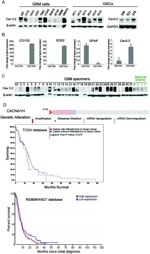
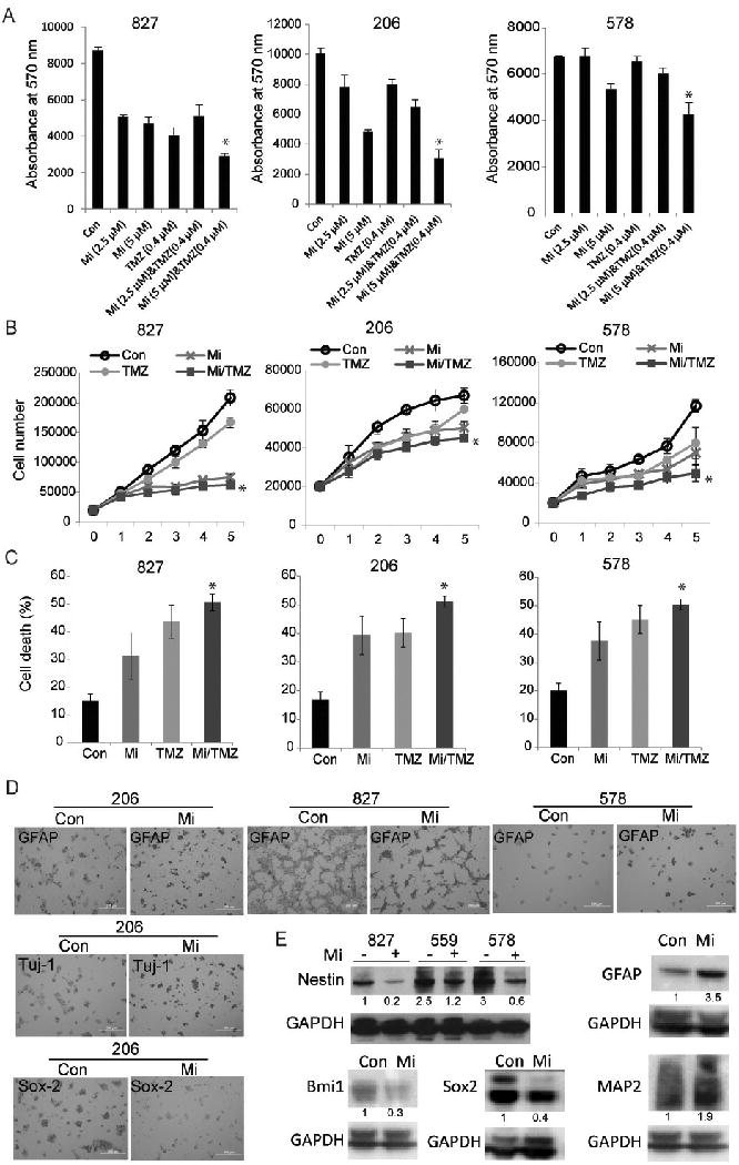
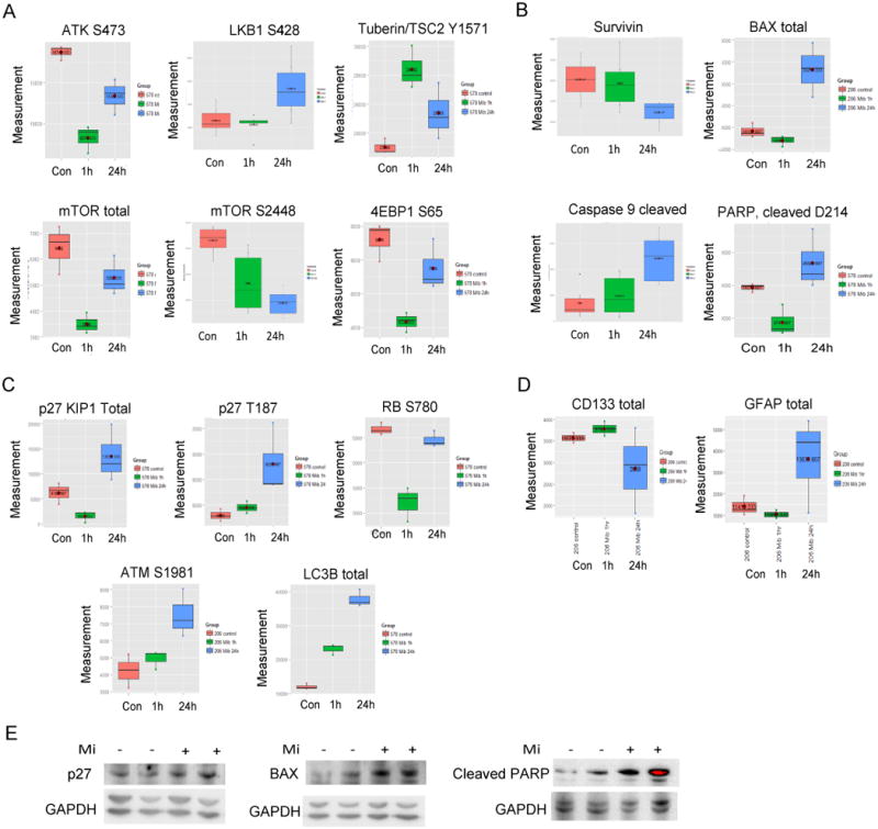
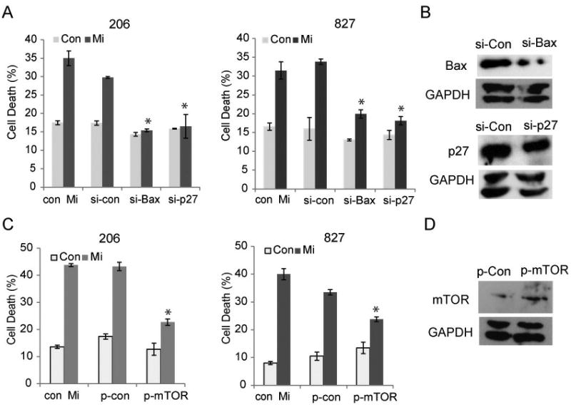
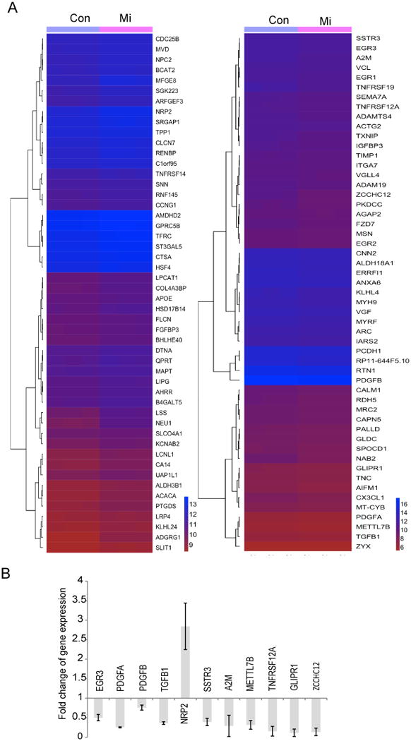
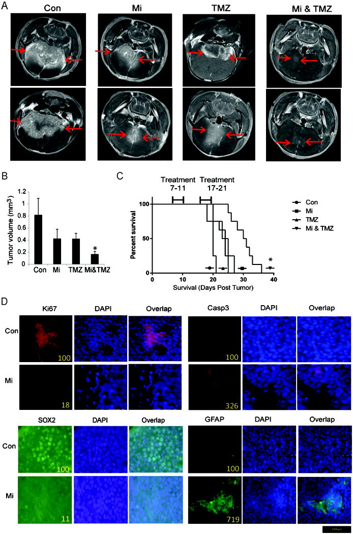
Glioblastoma (GBM) stem-like cells (GSC) promote tumor initiation, progression, and therapeutic resistance. Here, we show how GSCs can be targeted by the FDA-approved drug mibefradil, which inhibits the T-type calcium channel Cav3.2. This calcium channel was highly expressed in human GBM specimens and enriched in GSCs. Analyses of the The Cancer Genome Atlas and REMBRANDT databases confirmed upregulation of Cav3.2 in a subset of tumors and showed that overexpression associated with worse prognosis. Mibefradil treatment or RNAi-mediated attenuation of Cav3.2 was sufficient to inhibit the growth, survival, and stemness of GSCs and also sensitized them to temozolomide chemotherapy. Proteomic and transcriptomic analyses revealed that Cav3.2 inhibition altered cancer signaling pathways and gene transcription. Cav3.2 inhibition suppressed GSC growth in part by inhibiting prosurvival AKT/mTOR pathways and stimulating proapoptotic survivin and BAX pathways. Furthermore, Cav3.2 inhibition decreased expression of oncogenes (PDGFA, PDGFB, and TGFB1) and increased expression of tumor suppressor genes (TNFRSF14 and HSD17B14). Oral administration of mibefradil inhibited growth of GSC-derived GBM murine xenografts, prolonged host survival, and sensitized tumors to temozolomide treatment. Our results offer a comprehensive characterization of Cav3.2 in GBM tumors and GSCs and provide a preclinical proof of concept for repurposing mibefradil as a mechanism-based treatment strategy for GBM. Cancer Res; 77(13); 3479-90. ©2017 AACR.


Conflict of interest statement

Conflict of interest: Lloyd Gray is a paid consultant of Cavion LLC, who is commercializing the drug mibefradil.

Figures

Figure 1
Figure 1. Cav3.2 expression in Glioblastoma and correlation with stemness and patient survival
A) The glioblastoma cell lines U87, A172, U373, U251, T98G, U1242, SNB-19, SF-767, primary GBM cells GBM-6 and GBM-10, GBM stem cells XO-1, 2, 3, 4, 8, 9 and 206, 827 and 578 were lysed and immunoblotted for Cav3.2 and/or β-actin/GAPDH loading controls. B) Primary GBM cells (other than the ones of Fig. 1A) were sorted for CD133 expression by FACS and subjected to quantitative RT-PCR to determine the expression of CD133, SOX2, GFAP and Cav3.2. C) GBM human specimens G1-G31 and normal brains N1-4 were subjected to immunoblotting for Cav3.2 and β-Actin. D) TCGA (upper panel) and REMBRANDT (lower panel) data analyses of Cav3.2 (CACNA1H) mRNA expression and correlation with patient survival, Provisional, mRNA Expression z-Scores (RNA Seq V2 RSEM, with a z0score threshold 1.0) in upper panel. The analyses showed worse survival with high expression of Cav3.2. The above data show high expression of Cav3.2 in a subset of GBM and GSC and a trend towards inverse correlation with patient survival.
Figure 2
Figure 2. Cav3.2 blocker mibefradil inhibits GSC growth and enhances the effects of temozolomide in GSCs
A) GSCs 827, 206, 578 were treated with mibefradil (Mi) and/or temozolomide (TMZ) for 48 h. The cells were subsequently assessed for cell growth by Alamar blue assay. B) GSCs were treated with mibefradil and TMZ or control. The cells were subsequently assessed for proliferation by cell counting over a period of 5 days and growth curves were established. C) GSCs were treated with mibefradil and TMZ or control for 48 h and cell death was assessed by trypan blue assay. These data show that mibefradil induces GSC cell death, which is further enhanced by combinational treatment with TMZ (p<0.05). D) GSCs were seeded in pre-coated dishes with poly-L-ornithine. The cells were treated with mibefradil for 48 h, fixed and immunostained with differentiation markers, GFAP and Tuj-1 and stem cell marker Sox2. E) The GSCs were treated with mibefradil for 48 h then subjected to immunoblotting (quantified: numbers under blots) for the stem cell markers Nestin, Bmi1, Sox2 and astrocyte and neuronal markers GFAP and MAP2 and GAPDH control. These data show that mibefradil induces stem cell differentiation evidenced by the downregulation of Nestin Bmi1 and Sox2 and upregulation of GFAP, Tuj1 or MAP2. *, p<0.05 (Mi vs. combination).
Figure 3
Figure 3. Silencing of Cav3.2 induces GSC death and inhibits GSC growth
A) GSCs 827 and 206 were transfected with sh-Cav3.2 or sh-control for 48 h and subjected to immunoblotting for Cav3.2 and β-Actin. B) GSCs were either transfected with sh-Cav3.2 or sh-control for 48 h or treated with mibefradil for 48 h. The cells were subsequently assessed for proliferation by cell counting over a period of 5 days and growth curves were established. C) Cell death was assessed by trypan blue assay. These data show that silencing Cav3.2 inhibits GSC proliferation and induces cell death in a similar manner to mibefradil. *, p<0.05.
Figure 4
Figure 4. Mibefradil inhibits several oncogenic pathways in GSC
GSCs were treated with vehicle control (in red) or mibefradil for 1 h (in green) or 24 h (in blue) and the cell lysate was subjected to RPPA. Mibefradil downregulated A) the AKT/mTOR pathway whilst simultaneously upregulating LKB1 and TSC2, B) Mibefradil downregulated survivin and upregulated BAX, cleaved caspase 9 and cleaved PARP, C) Mibefradil upregulated p27, ATM and LC3B, D) Mibefradil downregulated CD133 and upregulated GFAP. E) RPPA verification by immunoblotting. These data show that mibefradil inhibits pro-survival pathways whilst inducing cell cycle arrest, apoptosis and DNA damage (p<0.05).
Figure 5
Figure 5. Mibefradil induces apoptosis via Bax, p27 and mTOR
A) GSCs 206 and 827 cells were transiently transfected with either si-control, si-p27 or si-Bax 48 h then treated with mibefradil or control for 48 h. Cell death was then assessed by trypan blue as previously described. Inhibition of either BAX or p27 abrogated mibefradil-induced cell death. These data show mibefradil acts to induce GSC cell death through the induction of apoptosis. B) Immunoblotting was undertaken to verify silencing of BAX and p27. C) GSCs were transfected with either control plasmid (p-con) or plasmid encoding mTOR (p-mTOR) for 48 h then treated with mibefradil or control for 48 h. Cell death was assessed by trypan blue assay. The data show mibefradil induces GSC cell death partly by inhibiting mTOR. D) Immunoblotting was undertaken to verify overexpression of mTOR. *, p<0.05.
Figure 6
Figure 6. Mibefradil alters gene expression in GSCs
GSCs (827) were treated with mibefradil or control for 24 h prior to total RNA extraction. RNA deep sequencing (RNA-seq) was performed. The data analysis (A) shows blockage of Cav3.2 by mibefradil alters gene expression, including downregulation of several oncogenes such as PDGFA, PDGFB, TGFB1, METTL7B, EGR3 and TNFRSF12A in GSC 827 and upregulation of tumor suppressive NRP2. B) Confirmation of RNA-seq data by quantitative PCR (p < 0.05 for all shown genes).
Figure 7
Figure 7. Mibefradil inhibits GSC xenograft growth and prolongs animal survival, also in combination with TMZ
A) GSC 827 cells were stereotactically implanted in the striatum of immunodeficient mice (n=10). Mibefradil or vehicle control were administered by daily oral gavage starting six days post-tumor implantation. The animals were subjected to MRI scan at 3 weeks after tumor implantation and B) tumor volumes were quantified. C) Animals were treated as in (A) and survival was analyzed. The data show that mibefradil significantly inhibits tumor growth and sensitizes tumors to TMZ treatment. D) Immunohistochemical staining of xenograft sections from (A) for the proliferation marker Ki67, the apoptotic marker cleaved-Caspase 3, stem cell marker Sox2 and astrocyte marker GFAP showing significantly reduced Ki67 and increased cleaved-caspase 3, as well as reduced Sox2 and elevated GFAP level in Mibefradil-treated xenografts (Sections at 40× magnification; Staining was quantified on sections with control set at 100). *, p<0.05.

References

    1. Wen PY, Reardon DA. Neuro-oncology in 2015: Progress in glioma diagnosis, classification and treatment. Nature reviews Neurology. 2016;12:69–70. - PubMed
    1. Galli R, Binda E, Orfanelli U, Cipelletti B, Gritti A, De Vitis S, Fiocco R, Foroni C, Dimeco F, Vescovi A. Isolation and characterization of tumorigenic, stem-like neural precursors from human glioblastoma. Cancer research. 2004;64:7011–21. - PubMed
    1. Singh SK, Hawkins C, Clarke ID, Squire JA, Bayani J, Hide T, Henkelman RM, Cusimano MD, Dirks PB. Identification of human brain tumour initiating cells. Nature. 2004;432:396–401. - PubMed
    1. Lathia JD, Mack SC, Mulkearns-Hubert EE, Valentim CL, Rich JN. Cancer stem cells in glioblastoma. Genes Dev. 2015;29:1203–17. - PMC - PubMed
    1. Bao S, Wu Q, McLendon RE, Hao Y, Shi Q, Hjelmeland AB, Dewhirst MW, Bigner DD, Rich JN. Glioma stem cells promote radioresistance by preferential activation of the DNA damage response. Nature. 2006;444:756–60. - PubMed

Publication types

Substances