. 2017 Sep;15(9):1138-1152.
doi: 10.1158/1541-7786.MCR-17-0003. Epub 2017 May 16.

Infiltrating Myeloid Cells Exert Protumorigenic Actions via Neutrophil Elastase

Affiliations

Infiltrating Myeloid Cells Exert Protumorigenic Actions via Neutrophil Elastase

Irina Lerman et al. Mol Cancer Res. 2017 Sep.

Abstract

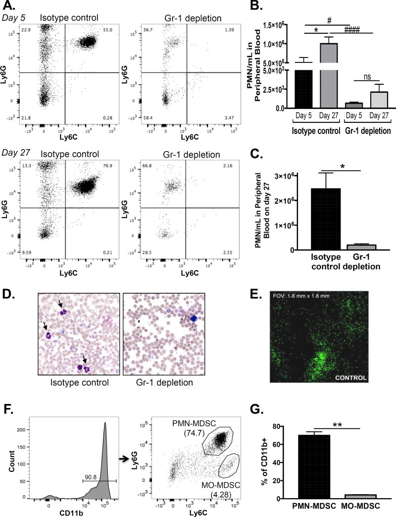
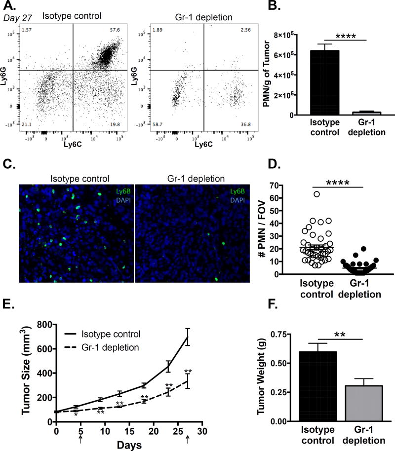
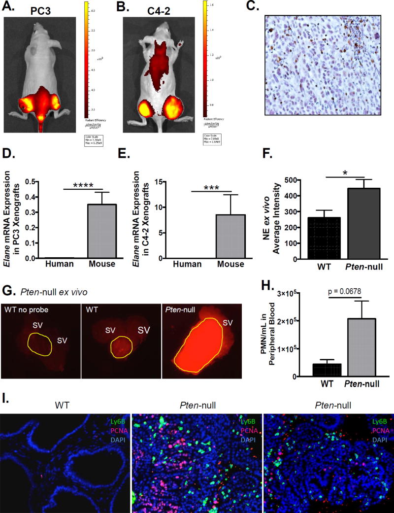
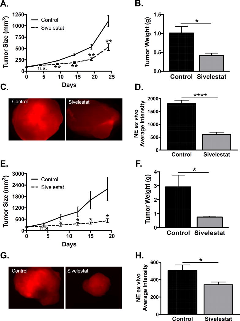
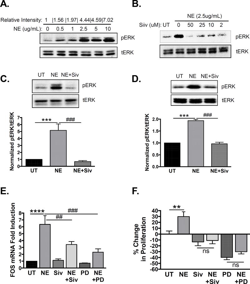
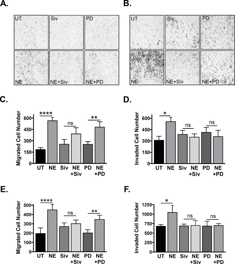
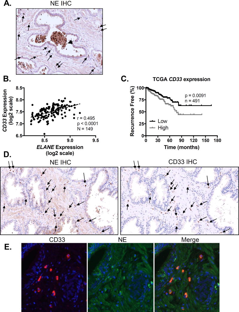
Tissue infiltration and elevated peripheral circulation of granulocytic myeloid-derived cells is associated with poor outcomes in prostate cancer and other malignancies. Although myeloid-derived cells have the ability to suppress T-cell function, little is known about the direct impact of these innate cells on prostate tumor growth. Here, it is reported that granulocytic myeloid-derived suppressor cells (MDSC) are the predominant tumor-infiltrating cells in prostate cancer xenografts established in athymic nude mice. MDSCs significantly increased in number in the peripheral circulation as a function of xenograft growth and were successfully depleted in vivo by Gr-1 antibody treatment. Importantly, MDSC depletion significantly decreased xenograft growth. We hypothesized that granulocytic MDSCs might exert their protumorigenic actions in part through neutrophil elastase (ELANE), a serine protease released upon granulocyte activation. Indeed, it was determined that NE is expressed by infiltrating immune cells and is enzymatically active in prostate cancer xenografts and in prostate tumors of prostate-specific Pten-null mice. Importantly, treatment with sivelestat, a small-molecule inhibitor specific for NE, significantly decreased xenograft growth, recapitulating the phenotype of Gr-1 MDSC depletion. Mechanistically, NE activated MAPK signaling and induced MAPK-dependent transcription of the proliferative gene cFOS in prostate cancer cells. Functionally, NE stimulated proliferation, migration, and invasion of prostate cancer cells in vitro IHC on human prostate cancer clinical biopsies revealed coexpression of NE and infiltrating CD33+ MDSCs.Implications: This report suggests that MDSCs and NE are physiologically important mediators of prostate cancer progression and may serve as potential biomarkers and therapeutic targets. Mol Cancer Res; 15(9); 1138-52. ©2017 AACR.


Figures

Figure 1
Figure 1. Granulocytic MDSCs expand in peripheral blood and infiltrate human prostate cancer xenografts
A. Peripheral blood MDSCs were assessed by flow cytometry in PC3 xenograft bearing nude mice on days 5 and 27 after initiation of isotype or Gr-1 antibody treatment. Representative plots of Ly6G vs. Ly6C expression, gated on CD11b+ myeloid cells, are shown (n=4/treatment group). B. The number of Ly6G+/Ly6C+ cells in Gr-1 depleted mice was compared to isotype controls on days 5 and 27 using one-way ANOVA with Bonferroni post-hoc testing (#compares day 5 numbers, p < 0.05, ####compares day 27 numbers, p < 0.0001). Isotype control and Gr-1 depletion groups on day 27 were compared to day 5 using one-way ANOVA with Bonferroni post-hoc testing (*compares days 5 and 27 control, p < 0.05, ns = not significant between days 5 and 27 depletion). C. The number of peripheral blood granulocytes on day 27 by automated cell counting was compared between isotype control and Gr-1 depletion mice using unpaired two-tailed t-test (n=4/treatment group; *p < 0.05). D. Representative Wright-Giemsa stained peripheral blood smears of isotype control and Gr-1 depletion mice on day 27. Arrows are cells with polymorphonuclear morphology. E. Whole mount immunofluorescence for Gr-1 in an untreated PC3 xenograft. F. Tumor infiltrating MDSCs were assessed by flow cytometry in control PC3 xenografts. Representative plot of Ly6G vs. Ly6C expression, gated on the CD11b+ myeloid population, is shown. Granulocytic (PMN; Ly6GHigh/Ly6CHigh) and monocytic (MO; Ly6GLow/Ly6CHigh) populations are indicated. G. The number of granulocytic and monocytic MDSCs was compared using paired two-tailed t-test (n=3; **p < 0.01).
Figure 2
Figure 2. Depletion of MDSCs with Gr-1 antibody suppresses human prostate cancer xenograft growth
A. Tumor infiltrating MDSCs were assessed by flow cytometry in PC3 xenograft bearing nude mice on day 27 after initiation of isotype or Gr-1 antibody treatment. Representative plots of Ly6G versus Ly6C expression, gated on the CD11b+ myeloid population, are shown (n=8/treatment group). B. The number of infiltrating granulocytic MDSCs (Ly6GHigh/Ly6CHigh) was compared between isotype control and Gr-1 depletion tumors, normalized to tumor weight, using unpaired two-tailed t-test (n=8; ****p < 0.0001). C. Representative immunofluorescence stain for Ly6B+ infiltrating granulocytic MDSCs in isotype control and Gr-1 depletion tumors is shown. D. The number of Ly6B+ cells was compared between isotype control and Gr-1 depletion tumors using unpaired two-tailed t-test (n=40/treatment group, 5 fields of view/8 tumors; ****p < 0.0001). E. Tumor size (length × width2 × 0.5) was measured every 3–4 days and compared between isotype control and Gr-1 depletion groups using unpaired two-tailed t-test (n=8 per treatment group; *p < 0.05, **p<0.01). Arrows indicate time points when MDSC depletion was assessed in peripheral blood. F. Tumor weight was compared between isotype control and Gr-1 depletion groups using unpaired two-tailed t-test (n=8 per treatment group; **p=0.01).
Figure 3
Figure 3. Infiltrating immune cells produce active neutrophil elastase in the prostate cancer microenvironment
Intra-tumoral neutrophil elastase activity was measured in-vivo using a NE specific optical probe in athymic nude mice bearing PC3 (A) and C4-2 (B) xenografts (n=3 per cell line). Representative IVIS images are shown. C. Immunohistochemistry of a representative PC3 xenograft using anti-NE antibody. Neutrophil elastase mRNA expression was quantified using quantitative PCR with human (ELANE) and mouse (Elane) specific primers for PC3 (D) and C4-2 (E) xenografts. Values were normalized to GAPDH and Gapdh mRNA expression, respectively. mRNA expression of mouse derived and human derived NE was compared using paired Wilcoxon signed-rank test (n=23 for PC3 and n=12 for C4-2; ****p<0.0001, *** p<0.001). F. Intra-prostatic NE activity in the prostatic ROI was quantified using ImageJ and compared between Pten-null and WT mice using unpaired two-tailed t-test (n=4 for each genotype; *p<0.05). G. Intra-prostatic NE activity was measured ex-vivo following IV administration of a NE specific optical probe to Pten-null and wild-type (WT) mice. First panel from a WT mouse that received no probe. Yellow outline indicates prostatic region of interest. SV = seminal vesicle. H. Peripheral blood MDSCs were assessed using flow cytometry in Pten-null and wild-type (WT) mice. The number of Ly6G+/Ly6C+ cells was compared using unpaired two-tailed t-test (n = 3 for each genotype). I. Representative immunofluorescence stains for Ly6B+ infiltrating granulocytic MDSCs and PCNA positive proliferating epithelium in WT and two different Pten-null prostates is shown (n=3 total for each group).
Figure 4
Figure 4. Inhibition of neutrophil elastase activity suppresses human prostate cancer xenograft growth
A. PC3 tumor size (length × width2 × 0.5) was measured every 3–4 days and compared between vehicle control and sivelestat groups using unpaired two-tailed t-test. B. PC3 tumor weight was compared between vehicle control and sivelestat groups using unpaired two-tailed t-test (n=4 per treatment group; *p<0.05, **p<0.01). C. PC3 intra-tumoral neutrophil elastase activity was measured ex-vivo using an NE specific optical probe. D. PC3 intra-tumoral NE activity was quantified using ImageJ and compared between vehicle control and sivelestat groups using unpaired two-tailed t-test (n=8 for vehicle control, n=6 for sivelestat; ****p<0.0001). E. C4-2 tumor size was measured every 3–4 days and compared between vehicle control and sivelestat groups using unpaired two-tailed t-test. F. C4-2 tumor weight was compared between vehicle control and sivelestat groups using unpaired two-tailed t-test (n=5 for control, n=6 for sivelestat; *p<0.05). G. C4-2 intra-tumoral neutrophil elastase activity was measured ex-vivo using an NE specific optical probe. H. C4-2 intra-tumoral NE activity was quantified using ImageJ and compared between vehicle control and sivelestat groups using unpaired two-tailed t-test (n=7 for vehicle control, n=7 for sivelestat; *p<0.05).
Figure 5
Figure 5. Neutrophil elastase activates MAPK signaling and induces MAPK-dependent gene transcription and proliferation in human prostate cancer cells
A. PC3 cells were serum starved and treated with increasing concentrations of NE for 15 minutes. pERK1/2 and tERK1/2 levels were examined by Western blot. Band densitometry of pERK1/2 to tERK1/2 was performed using ImageJ and normalized to untreated (0). B. PC3 cells were serum starved and treated with NE (2.5µg/mL) for 15 minutes in the presence of increasing concentrations of sivelestat. pERK1/2 and tERK1/2 levels were examined by Western blot. PC3 (C) or C4-2 (D) cells were serum starved and treated with NE (2.5µg/mL) for 15 minutes in the presence of sivelestat (2µM) or vehicle. pERK1/2 and tERK1/2 levels were examined by Western blot, and band densitometry performed using ImageJ and normalized to untreated (UT) samples. Multiple comparisons were performed using row matched one-way ANOVA with Dunnett’s post-hoc testing (n=6 for PC3 and n=3 for C4-2; ***p<0.001, ###p<0.001). E. C4-2 cells were serum starved and treated with NE (2.5µg/mL) for 6 hours in the presence of sivelestat (2µM), PD0325901 (50nM), or vehicle. cFOS mRNA expression was determined using quantitative PCR and normalized to GAPDH. Data were normalized to untreated samples and multiple comparisons were performed using row matched one-way ANOVA with Dunnett’s post-hoc testing (n=4; ****p<0.0001, ##p<0.01, ###p<0.001). F. C4-2 cells were serum starved and treated with NE (2.5µg/mL) for 24 hours in the presence of sivelestat (2µM), PD0325901 (50nM), or vehicle. Proliferation was examined by BrdU incorporation, and data were plotted as percent change in proliferation relative to untreated samples. Multiple comparisons were performed using ordinary one-way ANOVA with Bonferroni’s post-hoc testing (n=9; **p<0.01, ns=not significant).
Figure 6
Figure 6. Neutrophil elastase induces migration and invasion in human prostate cancer cells
A. C4-2 cells were serum starved and transferred into the upper chambers of 8um uncoated transwells in the presence of NE (2.5 ug/mL), sivelestat (2 uM), PD0325901 (50 nM), or vehicle. Cells were allowed to migrate for 24 hours towards a chemotactic gradient of 10% FBS. B. C4-2 cells were serum starved and transferred into the upper chambers of 8um Matrigel coated transwells in the presence of NE (2.5 ug/mL), sivelestat (2 uM), PD0325901 (50 nM), or vehicle. Cells were allowed to invade for 24 hours towards a chemotactic gradient of 10% FBS. C. Number of migrated C4-2 cells was quantified using ImageJ. Multiple comparisons were performed using row matched one-way ANOVA with Bonferroni’s post-hoc testing (n = 4; **** p < 0.0001, ** p < 0.01, ns = not significant). D. Number of invaded C4-2 cells was quantified using ImageJ (n = 4; * p < 0.05, ns = not significant). E. Number of migrated PC3 cells was quantified using ImageJ (n = 3; **** p < 0.0001, ** p < 0.01, ns = not significant). F. Number of invaded PC3 cells was quantified using ImageJ (n = 3; * p < 0.05, ns = not significant).
Figure 7
Figure 7. Neutrophil elastase is co-expressed with infiltrating CD33+ MDSCs in human prostates
A. Neutrophil elastase protein expression was determined by immunohistochemistry on human prostate cancer microarrays (image representative of n = 3 patients). Arrows indicate infiltrating NE positive cells. Arrowhead indicates NE positive glandular deposits. B. Human prostate cancer CD33 and ELANE mRNA expression data were obtained from Taylor (37) and plotted to examine correlation using two-tailed Pearson correlation analysis. C. Kaplan-Meier plots for patients expressing CD33 above (high) and below (low) the median CD33 expression threshold from the TCGA Provisional prostate cancer dataset (downloaded from http://cancergenome.nih.gov/) were constructed. Differences in recurrence free survival were assessed using the log-rank (Mantel-Cox) test. D. NE and CD33 protein expression were determined by immunohistochemistry on consecutive human prostate cancer samples (images representative of n = 3 patients). Arrows indicate infiltrating cells double positive for NE and CD33. E. NE and CD33 protein co-localization in human prostates was confirmed by immunofluorescence.

References

    1. Strasner A, Karin M. Immune Infiltration and Prostate Cancer. Frontiers in oncology. 2015;5:128. - PMC - PubMed
    1. Gurel B, Lucia MS, Thompson IM, Jr, Goodman PJ, Tangen CM, Kristal AR, et al. Chronic inflammation in benign prostate tissue is associated with high-grade prostate cancer in the placebo arm of the prostate cancer prevention trial. Cancer epidemiology, biomarkers & prevention : a publication of the American Association for Cancer Research, cosponsored by the American Society of Preventive Oncology. 2014;23(5):847–56. - PMC - PubMed
    1. Sharma J, Gray KP, Harshman LC, Evan C, Nakabayashi M, Fichorova R, et al. Elevated IL-8, TNF-alpha, and MCP-1 in men with metastatic prostate cancer starting androgen-deprivation therapy (ADT) are associated with shorter time to castration-resistance and overall survival. The Prostate. 2014;74(8):820–8. - PubMed
    1. Chi N, Tan Z, Ma K, Bao L, Yun Z. Increased circulating myeloid-derived suppressor cells correlate with cancer stages, interleukin-8 and −6 in prostate cancer. International journal of clinical and experimental medicine. 2014;7(10):3181–92. - PMC - PubMed
    1. Shafique K, Proctor MJ, McMillan DC, Qureshi K, Leung H, Morrison DS. Systemic inflammation and survival of patients with prostate cancer: evidence from the Glasgow Inflammation Outcome Study. Prostate cancer and prostatic diseases. 2012;15(2):195–201. - PubMed

Publication types

Substances