. 2017 Jul 18;8(29):47632-47641.
doi: 10.18632/oncotarget.17425.

Immune consequences of penfluridol treatment associated with inhibition of glioblastoma tumor growth

Affiliations

Immune consequences of penfluridol treatment associated with inhibition of glioblastoma tumor growth

Alok Ranjan et al. Oncotarget. .

Abstract

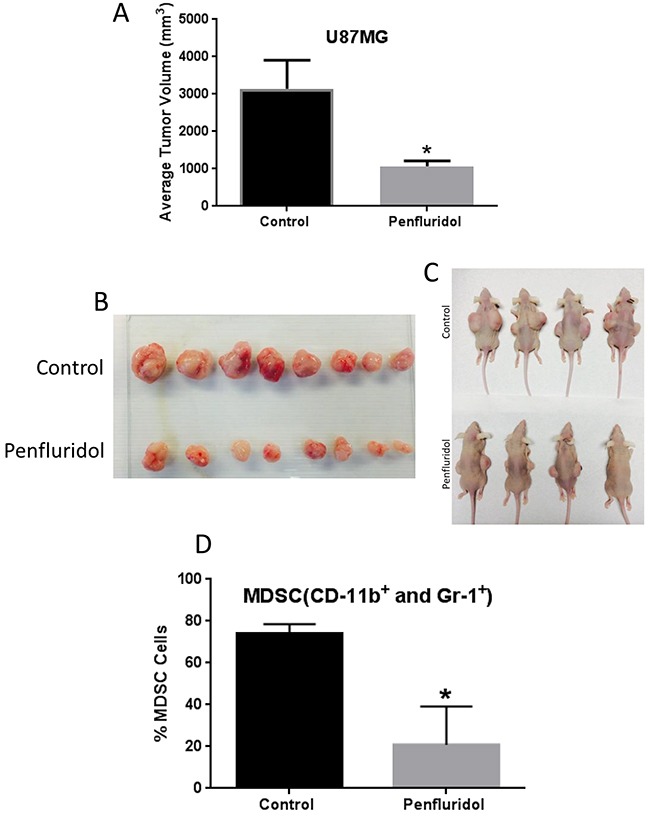
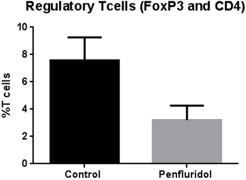
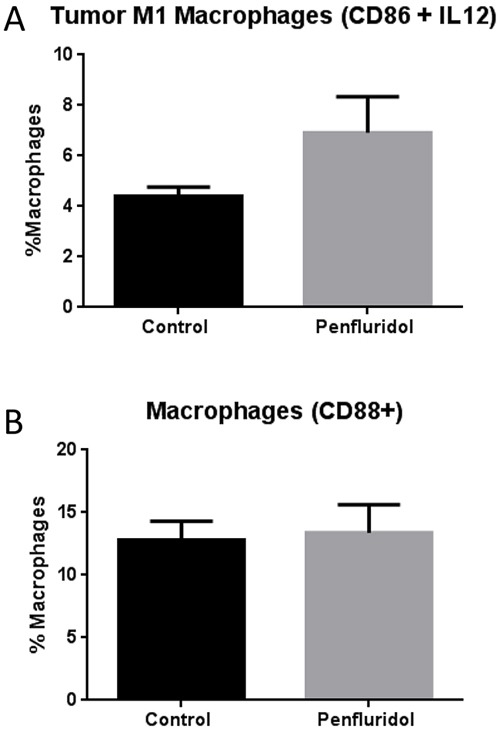
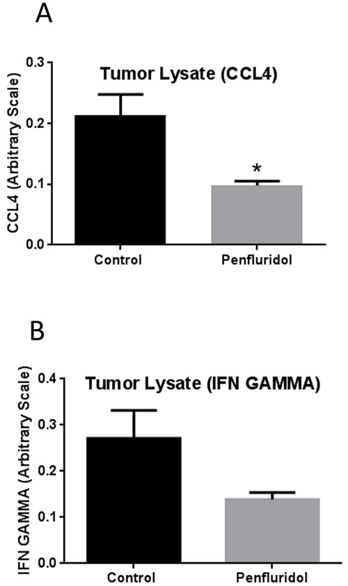
Glioblastoma is the most common and lethal brain tumor associated with only 12% median survival rate of patients. Despite the development of advanced surgical, radiation or use of combinations of anti-cancer drugs, treatment for glioblastoma patients is still a challenge. The major contributing factor in glioblastoma progression and resistive nature is its ability to evade the immune surveillance. Hence, modulating the immune system in glioblastoma tumors could be an important strategy for anticancer therapeutics. Penfluridol, an antipsychotic drug has been shown to have anti-cancer properties in our recently published studies. The present study evaluates the immune response of penfluridol in glioblastoma tumors. Our results demonstrated that penfluridol treatment significantly suppressed glioblastoma tumor growth. Our current results demonstrated about 72% suppression of myeloid derived suppressor cells (MDSCs) with penfluridol treatment in mouse bearing U87MG glioblastoma tumors. MDSCs are known to increase regulatory T cells (Treg), which are immunosuppressive in nature and suppresses M1 macrophages that are tumor suppressive in nature. Our results also showed suppression of regulatory T cells as well as elevation of M1 macrophages with penfluridol treatment by 58% and 57% respectively. Decrease in CCL4 as well as IFNγ with penfluridol treatment was also observed indicating decrease in overall tumor inflammation. This is the first report demonstrating immune modulations by penfluridol treatment associated with glioblastoma tumor growth suppression prompting further investigation to establish penfluridol as a treatment option for glioblastoma patients.

Keywords: MDSC; Treg; anti-psychotic drug; glioblastoma; macrophages.


Conflict of interest statement

CONFLICTS OF INTEREST

The authors declare no conflicts of interest.

Figures

Figure 1
Figure 1. Penfluridol suppresses glioblastoma tumor growth and associated MDSCs
1×10⁶ U87MG cells in 1:1 mixture of PBS and matrigel were implanted in right and left flanks of 4-6 week old athymic nude mice. Treatment with 10mg/kg penfluridol by oral gavage everyday started once tumor size was 70-100mm3 after tumor cells injection till day 48. Tumors from all control and penfluridol treated mice were removed at day 48. (A) Bar graph representing average tumor volume of control and penfluridol treated mice at day 48. Values were plotted as mean ± SEM. (B-C) Representative images of tumors as well as mice bearing tumors from control and treated group. Peripheral blood mononuclear cells (PBMCs) were collected from the blood obtained from control and penfluridol treated U87MG tumor-bearing athymic nude mice. Modulation of CD11b+ and Gr-1+ cells was analyzed by immunostaining and fluorescence cytometry to determine the effects of penfluridol on mouse myeloid derived suppressor cells (MDSCs); statistical analysis was performed by Student's t test. (D) Bar chart showing percent MDSCs which were double stained with CD11b and Gr-1 mouse antibodies. *Statistically significant at p<0.05 when compared with control.
Figure 2
Figure 2. Increase in spleen weight with penfluridol treatment: Spleens from control and penfluridol treated mice were removed and weights of spleens were taken
(A) Bar chart showing average weight of spleen in control and penfluridol treated group. Values were plotted as mean ± SEM. (B) Image of spleen from control and penfluridol treated mice.
Figure 3
Figure 3. Penfluridol suppresses regulatory T cells (Treg)
About 40 × 106 Human peripheral blood mononuclear cells (PBMCs) cells were injected i.p. in 100 μL PBS in SCID-NOD mice. The treatment group received 10mg/kg penfluridol by oral gavage everyday; control mice received vehicle alone. Modulation of regulatory T (Treg) cells was monitored by immunostaining and fluorescence cytometry to determine the effects of penfluridol. Values were plotted as mean ± SEM. Statistical analysis was performed by Student's t test.
Figure 4
Figure 4. Elevation of M1 macrophages in tumor with penfluridol treatment
Subcutaneously implanted U87MG glioblastoma tumors in SCID-NOD mice were removed from control and penfluridol treated mice at day 40. Tumors were processed and suspended into single cell suspension with the help of tumor dissociator kit and use of gentle MACS dissociator. Effect of penfluridol treatment on macrophages was analyzed by immunostaining and florescence cytometry. (A) Bar graph representing percent monocytic cells double stained with CD86 and IL12 human specific antibodies. (B) Percent cells which were single stained with CD88 human specific antibody.
Figure 5
Figure 5. Suppression of tumor inflammation with penfluridol treatment
After removing U87MG tumors, a part of the tumors were lysed and protein concentration was estimated in different control and penfluridol treated tumor lysate. Equal amount of protein was used to perform ELISA assay for CCL4 and IFNγ. (A-B) Bar graph representing CCL4 and IFNγ levels in the tumors from control and treatment group. Values were plotted as mean ± SEM. *Statistically significant at p<0.05 when compared with control.

References

    1. Zong H, Verhaak RG, Canoll P. The cellular origin for malignant glioma and prospects for clinical advancements. Expert Rev Mol Diagn. 2012;12:383–394. - PMC - PubMed
    1. Kohanbash G, Okada H. Myeloid-derived suppressor cells (MDSCs) in gliomas and glioma-development. Immunol Invest. 2012;41:658–679. - PubMed
    1. Raychaudhuri B, Rayman P, Ireland J, Ko J, Rini B, Borden EC, Garcia J, Vogelbaum MA, Finke J. Myeloid-derived suppressor cell accumulation and function in patients with newly diagnosed glioblastoma. Neuro Oncol. 2011;13:591–599. - PMC - PubMed
    1. Thaci B, Ahmed AU, Ulasov IV, Wainwright DA, Nigam P, Auffinger B, Tobias AL, Han Y, Zhang L, Moon KS, Lesniak MS. Depletion of myeloid-derived suppressor cells during interleukin-12 immunogene therapy does not confer a survival advantage in experimental malignant glioma. Cancer Gene Ther. 2014;21:38–44. - PMC - PubMed
    1. Kumar V, Patel S, Tcyganov E, Gabrilovich DI. The nature of myeloid-derived suppressor cells in the tumor microenvironment. Trends Immunol. 2016;37:208–220. - PMC - PubMed

MeSH terms