Review
. 2017 Aug;18(8):495-506.
doi: 10.1038/nrm.2017.48. Epub 2017 May 17.

Non-homologous DNA end joining and alternative pathways to double-strand break repair

Affiliations
Review

Non-homologous DNA end joining and alternative pathways to double-strand break repair

Howard H Y Chang et al. Nat Rev Mol Cell Biol. 2017 Aug.

Abstract

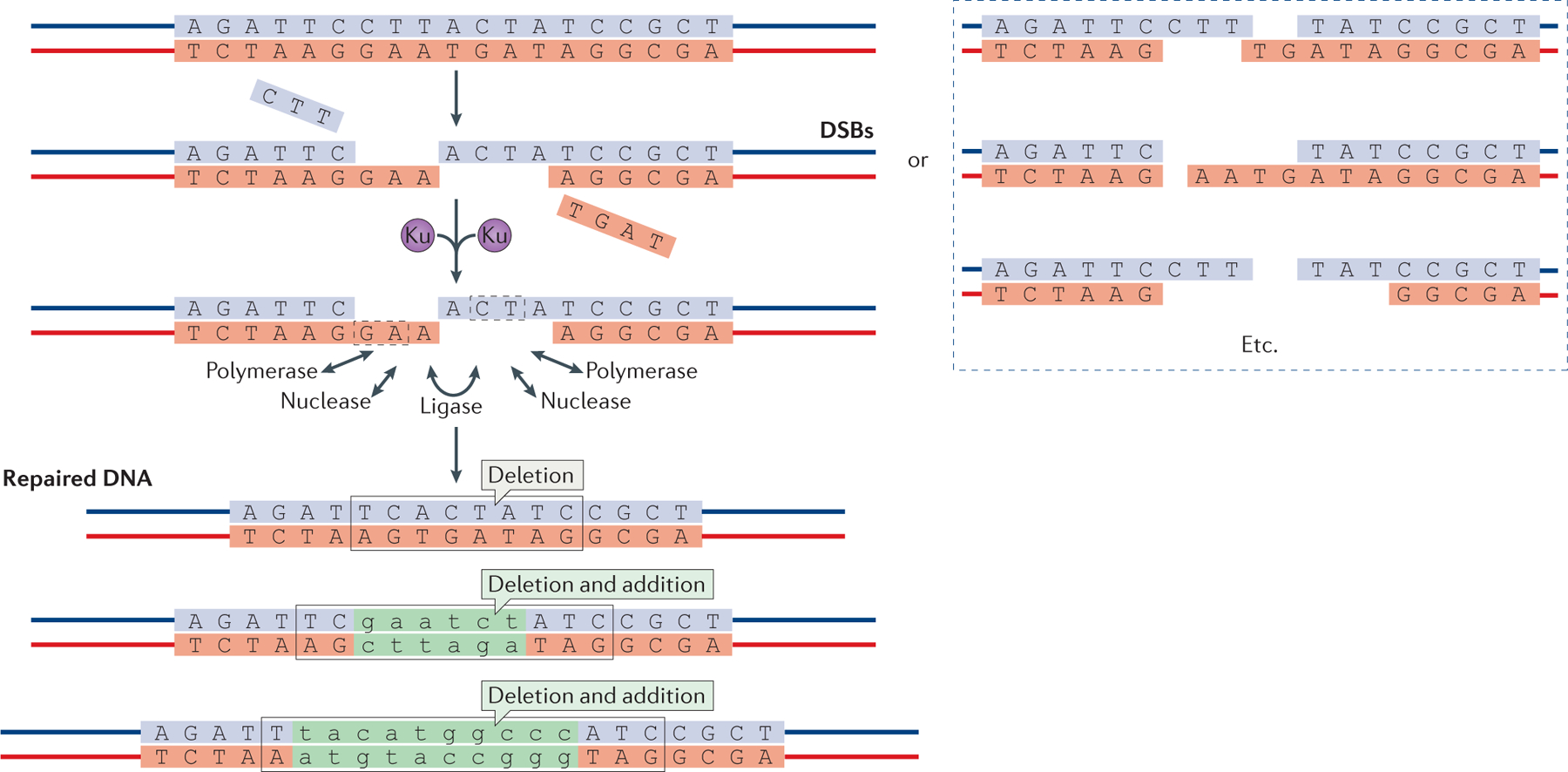
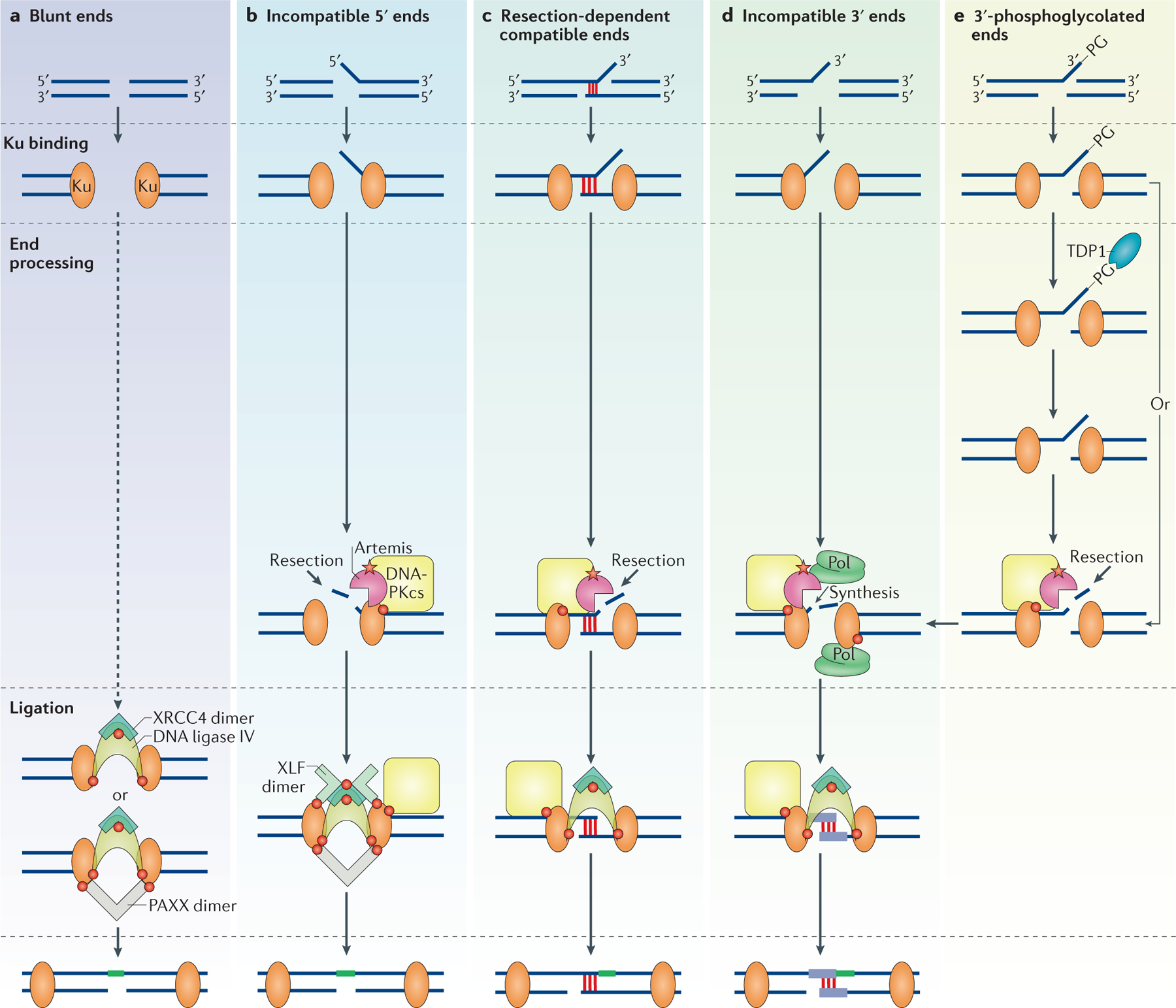
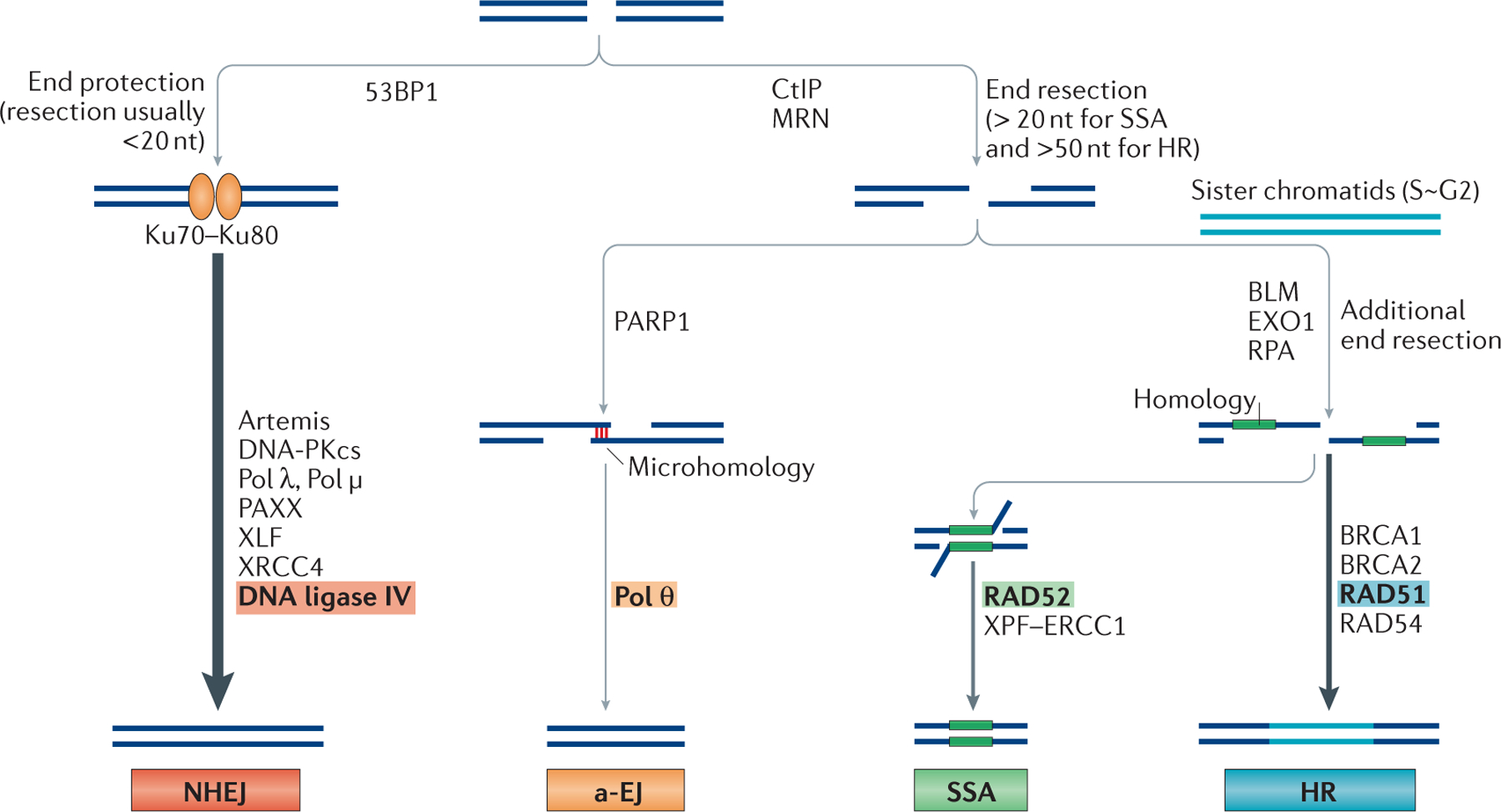
DNA double-strand breaks (DSBs) are the most dangerous type of DNA damage because they can result in the loss of large chromosomal regions. In all mammalian cells, DSBs that occur throughout the cell cycle are repaired predominantly by the non-homologous DNA end joining (NHEJ) pathway. Defects in NHEJ result in sensitivity to ionizing radiation and the ablation of lymphocytes. The NHEJ pathway utilizes proteins that recognize, resect, polymerize and ligate the DNA ends in a flexible manner. This flexibility permits NHEJ to function on a wide range of DNA-end configurations, with the resulting repaired DNA junctions often containing mutations. In this Review, we discuss the most recent findings regarding the relative involvement of the different NHEJ proteins in the repair of various DNA-end configurations. We also discuss the shunting of DNA-end repair to the auxiliary pathways of alternative end joining (a-EJ) or single-strand annealing (SSA) and the relevance of these different pathways to human disease.


Conflict of interest statement

Competing interests statement

The authors declare no competing interests.

Figures

Figure 1 |
Figure 1 |. Overview of non-homologous end joining.
Schematic of DNA double-strand breaks (DSBs) and their repair by non-homologous end joining (NHEJ) (top). The Ku70–Ku80 heterodimer binds to DSBs and improves their subsequent binding by the NHEJ polymerase, nuclease and ligase complexes. These enzymes can act on DSBs in any order to resect and add nucleotides. Multiple rounds of resection and addition are possible, and nuclease and polymerase activities at each of the two DNA ends seem to be independent. Microhomology between the two DNA ends, which is either already present (dashed boxes) or newly created when the polymerases add nucleotides in a template-independent manner, is often used to guide end joining. The process is error-prone and can result in diverse DNA sequences at the repair junction (bottom). However, NHEJ is also capable of joining two DNA ends without nucleotide loss from either DNA end and without any addition. Nucleotide additions are depicted in green lower case.
Figure 2 |
Figure 2 |. Non-homologous end joining proteins and their known interactions.
a | The non-homologous end joining (NHEJ) DNA protein kinase (DNA-PK) complex consists of a heterodimer of Ku70 and Ku80 plus DNA-PK catalytic subunit (DNA-PKcs). Ku70 and Ku80 consist of von Willebrand (vWA) domains, the Ku core and the nuclear localization sequence (NLS). Ku70 also contains a SAP (SAF-A/B, Acinus and PIAS) domain. DNA-PKcs consists of an amino-terminal domain with PQR and ABCDE autophosphorylation clusters implicated in its activation, a FAT (FRAP, ATM, TRRAP) domain, followed by the phosphatidylinositol 3-kinase (PI3K) domain, and the FAT-C (carboxy-terminal) domain. b | The NHEJ nucleases consist of Artemis and aprataxin and PNKP-like factor (APLF). Artemis has a catalytic β-lactamase domain, a cleavage and polyadenylation specificity factor (β-CASP) domain and a disordered C terminus. Amino acids 454–458 bind to aa 1–7 to auto-inhibit Artemis activity. APLF consists of a forkhead-associated (FHA) domain, a middle (MID) domain and the poly(ADP-ribose)-binding zinc-finger (PBZ) domain. c | The polymerases involved in NHEJ are Pol λ, Pol μ and terminal deoxynucleotidytransferase (TdT). They consist of a BRCA1 C terminus (BRCT) domain, a lyase domain and a nucleotidyltransferase domain. d | The DNA ligase complex consists of DNA ligase IV, X-ray repair cross-complementing protein 4 (XRCC4), XRCC4-like factor (XLF) and paralogue of XRCC4 and XLF (PAXX). DNA ligase IV consists of an N-terminal DNA binding domain, a catalytic core and an XRCC4 interaction domain (XID) flanked by the BRCT I and BRCT II domains. XRCC4, XLF and PAXX are structurally similar, with an N-terminal head domain, helical domain and a C terminus. Protein domains are shown in solid colour and linker regions in grey.
Figure 3 |
Figure 3 |. The various non-homologous end joining subpathways.
Various non-homologous end joining (NHEJ) proteins may associate at common NHEJ substrates. Red circles represent known protein–protein interactions depicted in FIG. 2. The red star represents the interaction between Artemis and DNA-dependent protein kinase catalytic subunit (DNA-PKcs), which results in the activation of the endonuclease activity of Artemis. The diagram depicts ligation (in green) only of the top strand, but this process will inevitably proceed to the bottom strand (Supplementary information S2 (figure)). a | Blunt DNA ends are preferentially repaired without end processing and their ligation can be stimulated by paralogue of XRCC4 and XLF (PAXX). b | Incompatible 5′ overhanging ends are preferentially processed with resection of the 5′ overhang by the Artemis–DNA-PKcs complex, followed by blunt-end ligation that is stimulated by XRCC4-like factor (XLF) and PAXX. c | Resection-dependent compatible ends that have a short stretch of microhomology (~4 nucleotides of base pairing) along with a non-base paired flap only require Artemis–DNA-PKcs to cleave off the flap for ligation to occur. d | Incompatible 3′ overhanging ends are processed by iterative events of end resection and nucleotide synthesis by DNA polymerases (Pol) to generate short regions of base pairing (purple) before ligation. e | 3′-phosphoglycolated (3′-PG) ends can form on recessed or blunt ends or on a DNA overhang, and can be processed by tyrosyl DNA phosphodiesterase 1 (TDP1). Alternatively, Artemis–DNA-PKcs can bypass the 3′-PG and probably other end modifications and can endonucleolytically resect the ends that contain the modifications. Ku, Ku70–Ku80; XRCC4, X-ray repair cross-complementing protein 4.
Figure 4 |
Figure 4 |. Double-strand break repair pathway choice.
DNA double-strand breaks (DSBs) can be repaired by the classical non-homologous end joining (NHEJ) pathway, the alternative end joining (a-EJ) pathway, the single-strand annealing (SSA) pathway or by homologous recombination (HR). The major differences in pathway choice are the requirement for substantial DNA end resection. p53-binding protein 1 (53BP1) is a chromatin remodeller and a positive regulator of NHEJ. Although the complex of Artemis and DNA-dependent protein kinase catalytic subunit (DNA-PKcs) can carry out some resection (typically <20 nucleotides), the NHEJ pathway does not require extensive end resection and the ends are mostly protected by the binding of Ku70–Ku80. By contrast, carboxy-terminal binding protein interacting protein (CtIP) and the MRN (MRE11–RAD50–NBS1 (Nijmegen breakage syndrome protein 1)) complex are involved in extensive 5′ to 3′ resection of regions of the duplex to generate stretches of single-strand DNA (ssDNA) at DNA ends for a-EJ, SSA and HR. SSA typically requires >20 bp of microhomology, whereas the requirement for a-EJ is <25 bp. Poly(ADP-ribose) polymerase 1 (PARP1) and DNA polymerase θ (Pol θ) are important for a-EJ. Bloom syndrome RecQ-like helicase (BLM) and exonuclease 1 (EXO1) provide additional resection, and replication protein A (RPA) binds to ssDNA to promote the SSA and the HR pathways. RAD52-mediated annealing of large regions of homology is key for the SSA pathway. The xeroderma pigmentosum group F (XPF)–ERCC1 complex cuts the remaining 3′ overhangs before ligation. By contrast, RAD51-mediated strand exchange and its association with BRCA1, BRCA2 and RAD54 are essential for promoting the HR pathway. PAXX, paralogue of XRCC4 and XLF; XLF, XRCC4-like factor; XRCC4, X-ray repair cross-complementing 4.
Figure 5 |
Figure 5 |. Microhomology length requirement of DNA-end joining pathways.
Non-homologous end joining (NHEJ) uses short stretches of microhomology (from 0 to 4 bp), whereas alternative end joining (a-EJ) uses <20 bp of microhomology (most commonly 4–6 bp). The single-strand annealing (SSA) pathway uses >20 bp of homology (for SSA, homology is a more appropriate term than microhomology); the relative use of SSA depends on the organism and the length of homology.

References

    1. Lieber MR The mechanism of double-strand DNA break repair by the nonhomologous DNA end-joining pathway. Annu. Rev. Biochem 79, 181–211 (2010). - PMC - PubMed
    1. Lieber MR & Karanjawala ZE Ageing, repetitive genomes and DNA damage. Nat. Rev. Mol. Cell Biol 5, 69–75 (2004). - PubMed
    1. Martin GM, Smith AC, Ketterer DJ, Ogburn CE & Disteche CM Increased chromosomal aberrations in first metaphases of cells isolated from the kidneys of aged mice. Isr. J. Med. Sci 21, 296–301 (1985). - PubMed
    1. Meek K, Dang V & Lees-Miller SP DNA-PK: the means to justify the ends? Adv. Immunol 99, 33–58 (2008). - PubMed
    1. Chang HHY et al. Different DNA end configurations dictate which NHEJ components are most important for joining efficiency. J. Biol. Chem 291, 24377–24389 (2016). - PMC - PubMed
    2. This paper describes a biochemical reconstitution of NHEJ using a direct gel assay (without PCR) and all of the major NHEJ components.

Publication types