

Homonymous Hemianopsia Due to Posterior Cortical Atrophy
- PMID: 28512507
- PMCID: PMC5417079
- DOI: 10.1080/01658107.2016.1278556
Homonymous Hemianopsia Due to Posterior Cortical Atrophy
Abstract
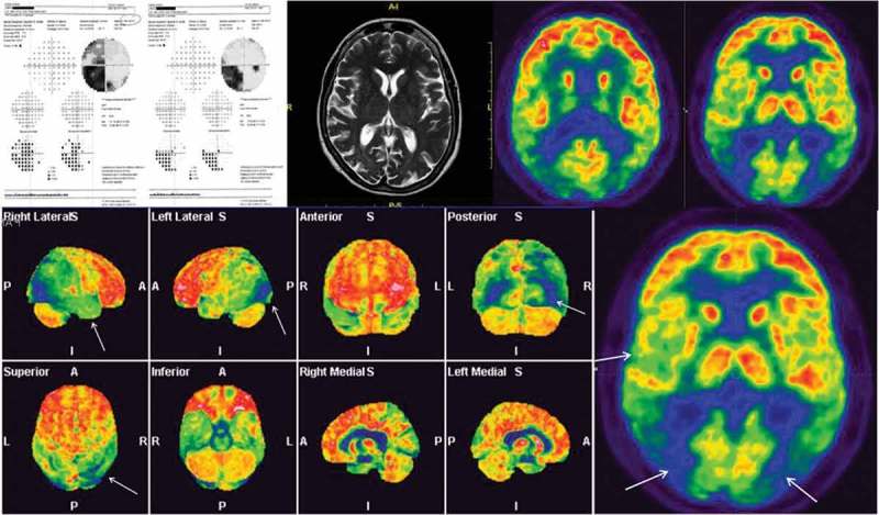
A 63-year-old woman presented to her ophthalmologist complaining of reading difficulties for two years. Ophthalmological examination revealed a homonymous hemianopsia. Brain magnetic resonance imaging (MRI) scan was interpreted as normal, but positron emission tomography (PET) showed areas of posterior brain hypometabolism. This case highlights the high diagnostic suspicion that ophthalmologists should have regarding posterior cortical atrophy (including the visual variant of Alzheimer disease) in patients complaining of reading difficulties in the setting of a normal ophthalmic examination.
Keywords: Brain PET; homonymous hemianopsia; posterior cortical atrophy; visual variant of Alzheimer disease.
Figures
Similar articles
-
The woman who needed a pet.Surv Ophthalmol. 2006 Nov-Dec;51(6):592-5. doi: 10.1016/j.survophthal.2006.08.002. Surv Ophthalmol. 2006. PMID: 17134649
-
[Homonymous hemianopia and posterior cortical atrophy].Rev Neurol (Paris). 2009 Mar;165(3):256-62. doi: 10.1016/j.neurol.2008.10.010. Epub 2009 Jan 4. Rev Neurol (Paris). 2009. PMID: 19124140 French.
-
Clinical Reasoning: An 80-Year-Old Woman With a Homonymous Hemianopsia.Neurology. 2022 Oct 18;99(16):713-717. doi: 10.1212/WNL.0000000000201175. Epub 2022 Aug 29. Neurology. 2022. PMID: 36038269
-
[Visual impairment in posterior cortical atrophy--visual variant of Alzheimer's disease in the ophthalmic practice].Klin Oczna. 2014;116(3):215-8. Klin Oczna. 2014. PMID: 25799788 Review. Polish.
-
[Homonymous hemianopsia and oral ovulation inhibitors. A neuro-ophthalmologic consideration].Dtsch Gesundheitsw. 1975 Mar 2;30(10):476-9. Dtsch Gesundheitsw. 1975. PMID: 1092537 Review. German.
Cited by
-
Alzheimer's visual variant: a report of a diagnosis easily missed on ophthalmic examination.Int J Ophthalmol. 2024 Aug 18;17(8):1571-1575. doi: 10.18240/ijo.2024.08.24. eCollection 2024. Int J Ophthalmol. 2024. PMID: 39156773 Free PMC article. No abstract available.
-
Posterior Type of Alzheimer's Dementia Presenting with Homonymous Hemianopsia.Dement Neurocogn Disord. 2017 Dec;16(4):128-131. doi: 10.12779/dnd.2017.16.4.128. Epub 2017 Dec 31. Dement Neurocogn Disord. 2017. PMID: 30906384 Free PMC article.
References
-
- Brazis PW, Lee AG, Graff-Radford N, Desai NP.. Homonymous visual field defects in patients without corresponding structural lesions on neuroimaging. J Neuroophthalmol 2000;20:92–96. - PubMed
-
- Benson DF, Davis RJ, Snyder BD.. Posterior cortical atrophy. Arch Neurol 1988;45:789–793. - PubMed
-
- Snowden JS, Stopford CL, Julien CL, Thompson JC, Davidson Y, Gibbons L, Pritchard A, Lendon CL, Richardson AM, Varma A, Neary D, Mann D.. Cognitive phenotypes in Alzheimer’s disease and genetic risk. Cortex 2007;43:835–845. - PubMed
-
- Zakzanis KK, Boulos MI.. Posterior cortical atrophy. Neurologist 2001;7:341–349. - PubMed
LinkOut - more resources
Full Text Sources
Other Literature Sources