Lhermitte-Duclos Disease and Cerebellar Gangliocytoma-An Incidental Finding in a Patient with Gradual Vision Loss
- PMID: 28512508
- PMCID: PMC5417168
- DOI: 10.1080/01658107.2017.1291685
Lhermitte-Duclos Disease and Cerebellar Gangliocytoma-An Incidental Finding in a Patient with Gradual Vision Loss
Abstract
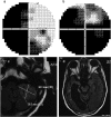
A 50-year-old male patient presented to the neuro-ophthalmology clinic with chief complaints of gradual decrease in vision in both eyes, more in the left eye, for 6 years. On general examination, the patient had a hemiplegic gait. His presenting acuity was 20/50 in the right eye and 20/320 in the left eye, not improving further. He had dense posterior subcapsular cataracts in both eyes, and fundus examination revealed pale discs. Humphrey visual field tests 30-2 revealed a vertical nasal midline defect in the right eye and grossly depressed fields in the left eye. Keeping in mind the above findings, the authors requested for a magnetic resonance imaging (MRI) of the brain. The brain MRI shows a large infarct in the right parieto-occipital lobe and a small circumscribed lesion in the left cerebellum. The radiologist opined that it could possibly be a gangliocytoma of the cerebellum, and a possible diagnosis of Lhermitte-Duclos syndrome was made.
Keywords: Lhermitte-Duclos disease; cerebellar gangliocytoma.
Figures
References
-
- Nowak DA, Trost HA.. Lhermitte-Duclos disease (dysplastic cerebellar gangliocytoma): a malformation, hamartoma or neoplasm? Acta Neurol Scand 2002;105:137–145. - PubMed
-
- Peltier J, Lok C, Fichten A, Bruniau A, Lefranc M, Toussaint P, Desenclos C, Le Gars D. Lhermitte-Duclos disease and Cowden’s syndrome. Report of two cases. Neuro-Chirurgie 2006;52:407–414 - PubMed
-
- Yagci-Kupeli B, Oguz KK, Bilen MA, Yalcin B, Akalan N, Buyukpamukcu M.. An unusual cause of posterior fossa mass: Lhermitte-Duclos disease. J Neurol Sci 2010;290:138–141. - PubMed
LinkOut - more resources
Full Text Sources
Other Literature Sources

