. 2017 May 17:8:15383.
doi: 10.1038/ncomms15383.

Crystal structure of a multi-domain human smoothened receptor in complex with a super stabilizing ligand

Affiliations

Crystal structure of a multi-domain human smoothened receptor in complex with a super stabilizing ligand

Xianjun Zhang et al. Nat Commun. .

Abstract

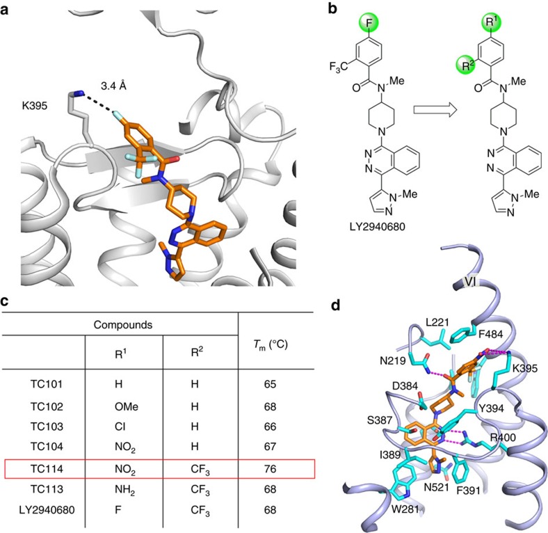
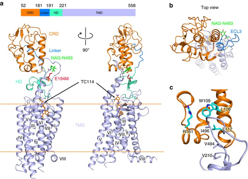
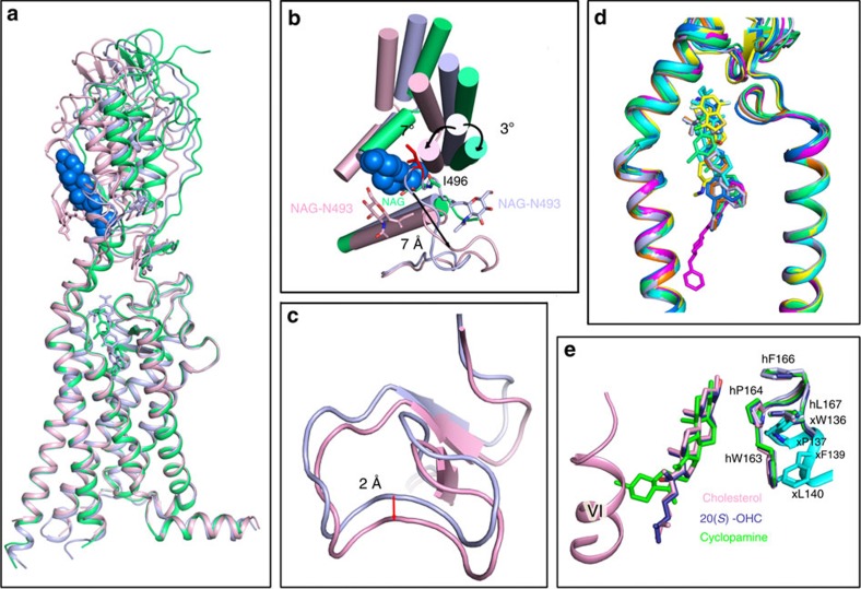
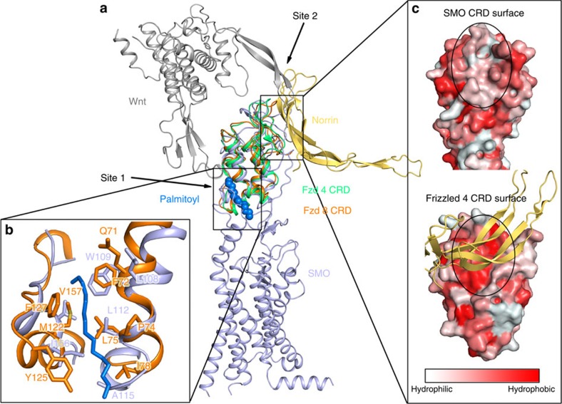
The Smoothened receptor (SMO) belongs to the Class Frizzled of the G protein-coupled receptor (GPCR) superfamily, constituting a key component of the Hedgehog signalling pathway. Here we report the crystal structure of the multi-domain human SMO, bound and stabilized by a designed tool ligand TC114, using an X-ray free-electron laser source at 2.9 Å. The structure reveals a precise arrangement of three distinct domains: a seven-transmembrane helices domain (TMD), a hinge domain (HD) and an intact extracellular cysteine-rich domain (CRD). This architecture enables allosteric interactions between the domains that are important for ligand recognition and receptor activation. By combining the structural data, molecular dynamics simulation, and hydrogen-deuterium-exchange analysis, we demonstrate that transmembrane helix VI, extracellular loop 3 and the HD play a central role in transmitting the signal employing a unique GPCR activation mechanism, distinct from other multi-domain GPCRs.


Conflict of interest statement

R.C.S. and F.X. are founders and R.C.S. is a board member of Bird Rock Bio, a company focused on GPCR therapeutic antibodies. The remaining authors declare no competing financial interests.

Figures

Figure 1
Figure 1. Design and synthesis of SMO ligand TC114 for crystallographic studies.
(a) Close-up view of the LY2940680 binding pocket in ΔCRD–SMO structure (PBD ID: 4JKV). (b) Design and evolution of LY2940680 analogues for crystallization study by variation of the substituents on aromatic ring. (c) The representative Tm values for LY2940680 and its analogues according to the CPM thermal shift assay. (d) Close-up view of TC114 binding pocket. TC114 (orange carbons) and SMO residues (cyan carbons) involved in ligand binding are shown in stick representation. The receptor is shown in light blue cartoon representation. Other elements are coloured as follows: oxygen, red; nitrogen, dark blue; sulfur, yellow. Hydrogen bonds are displayed as magenta dashed lines.
Figure 2
Figure 2. Overall structure of the multi-domain human SMO.
(a) Overall structure of the human SMO in complex with TC114 determined at an XFEL. TC114 is shown as orange sticks. The CRD, linker, HD and TMD are indicated as orange, marine, green cyan and light blue cartoons, respectively. The membrane boundary is labelled, as an orange dashed line. N-linked glycans (NAG) are shown in green sticks. (b) Top view of the SMO from the extracellular side. A hydrophobic pocket is formed by the CRD hydrophobic groove and ECL3 (marine loop). (c) Key residues in the CRD and ECL3 defining the hydrophobic pocket are shown in cyan and light blue sticks, respectively.
Figure 3
Figure 3. Unique multi-domain interaction and modularity in the human SMO.
(a) Superposition of three multi-domain SMO structures: SMO solved at XFEL, SMO in complex with cholesterol (PBD ID: 5L7D) and SMO in complex with vismodegib (PBD ID: 5L7I) are shown in light blue, pink and lime green cartoons, respectively. The cholesterol in the 5L7D structure is shown in marine spheres. (b) Top view of the extracellular side. The glycans from XFEL, cholesterol-bound and vismodegib-bound structures are shown in light blue, pink and lime green sticks, respectively. I496 from XFEL structure is shown in red sticks. (c) Hinge domains (HDs) from SMO in complex with TC114 (light blue) and SMO in complex with cholesterol (PBD ID: 5L7D; pink) are superimposed and shown as cartoons. (d) Close-up view of superimposed structures of the TMD bundle of SMO with different ligands bound. SMO in complex with TC114 (light blue), SMO in complex with vismodegib (PBD ID: 5L7I; lime green), ΔCRD–SMO in complex with LY2940680 (PBD ID: 4JKV; orange), ΔCRD–SMO in complex with Anta XV (PDB ID: 4QIM; marine), ΔCRD–SMO in complex with SAG 1.5 (PDB ID: 4QIN; yellow), ΔCRD–SMO in complex with SANT1 (PDB ID: 4N4W; magenta) and ΔCRD–SMO in complex with Cyclopamine (PDB ID: 4O9R; cyan) are superimposed and shown as cartoons. (e) Superimposed structures of SMO CRDs. CRD from multi-domain SMO structure solved at XFEL (light blue), CRD from multi-domain SMO structure in complex with cholesterol (PBD ID: 5L7D; pink), CRD from xSMO in the apo state (PBD ID: 5KZZ; cyan), CRD from xSMO in complex with 20(S)-OHC (PBD ID: 5KZV; deep blue) and CRD from xSMO in complex with cyclopamine (PBD ID: 5KZY; green) are shown in cylindrical helices. Key residues, cholesterol, 20(S)-OHC and cyclopamine are shown in sticks.
Figure 4
Figure 4. Comparison of the CRD of the human SMO with the Frizzled receptors.
(a) Side view of superimposed structures of the human SMO (hSMO) CRD with the hFzd-4 (PDB ID: 5CL1) and mFzd-8 CRDs (PDB ID: 4F0A). The SMO, hFzd-4 and mFzd-8 are shown as cartoons in light blue, lime green and orange, respectively. The Wnt and Norrin are shown as dark grey and yellow orange cartoons, respectively. The palmitoyl group in the mFzd-8 CRD is shown in marine spheres. (b) Site 1: Close-up view of the palmitoyl group with interacting residues as orange sticks. The palmitoyl group is shown in marine sticks. The residues from SMO forming the hydrophobic pocket are shown in light blue sticks, mFzd-8 in orange sticks. (c) Site 2: Surface of SMO and hFzd-4 CRD. Norrin is shown in light orange cartoons. The colour gradient from light red to dark red corresponds to the change of surface property from hydrophilic to hydrophobic. The Norrin binding site on hFzd-4 CRD surface is labelled by a black dashed circle with corresponding site also marked on the SMO surface.

Comment in

References

    1. Pasca di Magliano M. & Hebrok M. Hedgehog signalling in cancer formation and maintenance. Nat. Rev. Cancer 3, 903–911 (2003). - PubMed
    1. Zhao Y., Tong C. & Jiang J. Hedgehog regulates smoothened activity by inducing a conformational switch. Nature 450, 252–258 (2007). - PubMed
    1. Wang C. et al. Structure of the human smoothened receptor bound to an antitumour agent. Nature 497, 338–343 (2013). - PMC - PubMed
    1. Ingham P. W. & McMahon A. P. Hedgehog signaling in animal development: paradigms and principles. Genes Dev. 15, 3059–3087 (2001). - PubMed
    1. Zhao Z. et al. An essential role for Grk2 in Hedgehog signalling downstream of Smoothened. EMBO Rep. 17, 739–752 (2016). - PMC - PubMed

Publication types

LinkOut - more resources