. 2018 Jan;11(1):50-60.
doi: 10.1038/mi.2017.38. Epub 2017 May 17.

Jak3 deficiency blocks innate lymphoid cell development

Affiliations

Jak3 deficiency blocks innate lymphoid cell development

M L Robinette et al. Mucosal Immunol. 2018 Jan.

Abstract

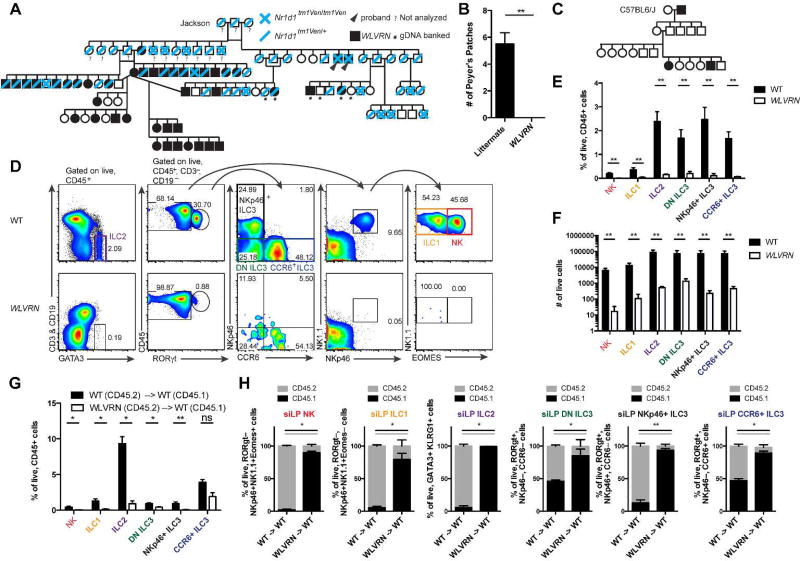
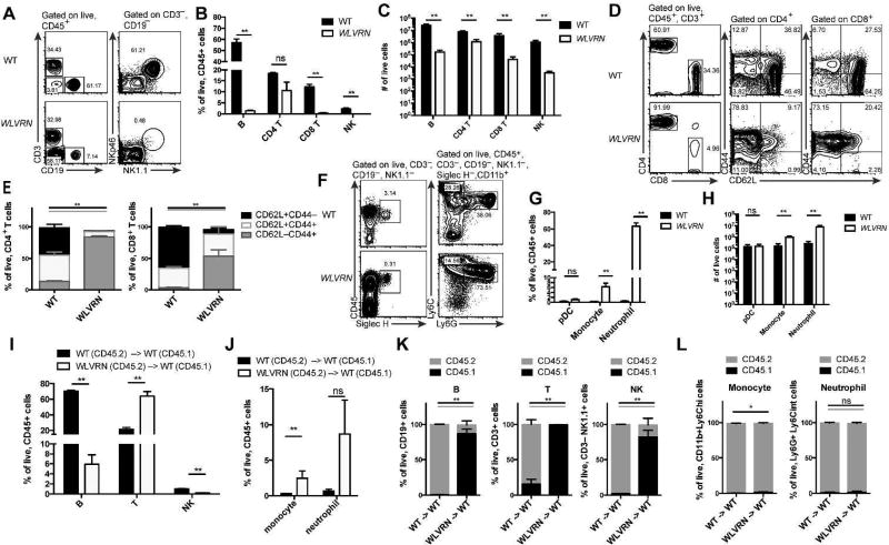
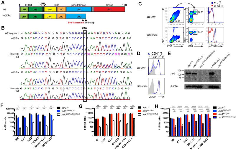
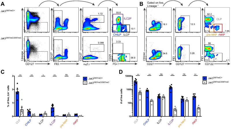
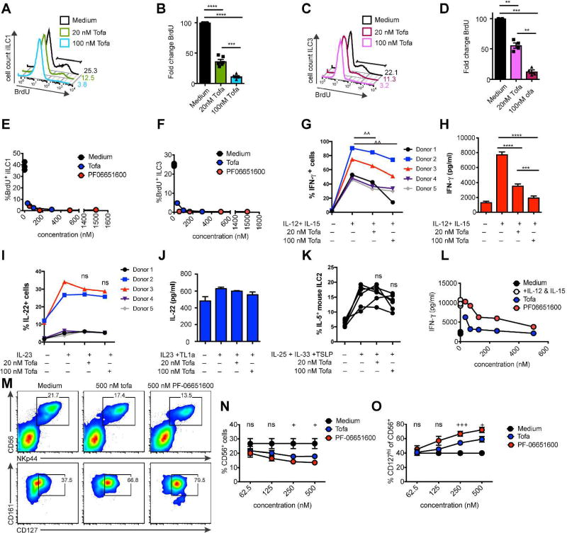
Loss-of-function mutations in the tyrosine kinase JAK3 cause autosomal recessive severe combined immunodeficiency (SCID). Defects in this form of SCID are restricted to the immune system, which led to the development of immunosuppressive JAK inhibitors. We find that the B6.Cg-Nr1d1tm1Ven/LazJ mouse line purchased from Jackson Laboratories harbors a spontaneous mutation in Jak3, generating a SCID phenotype and an inability to generate antigen-independent professional cytokine-producing innate lymphoid cells (ILCs). Mechanistically, Jak3 deficiency blocks ILC differentiation in the bone marrow at the ILC precursor and the pre-NK cell progenitor. We further demonstrate that the pan-JAK inhibitor tofacitinib and the specific JAK3 inhibitor PF-06651600 impair the ability of human intraepithelial ILC1 (iILC1) to produce IFN-γ, without affecting ILC3 production of IL-22. Both inhibitors impaired the proliferation of iILC1 and ILC3 and differentiation of human ILC in vitro. Tofacitinib is currently approved for the treatment of moderate-to-severely active rheumatoid arthritis. Both tofacitinib and PF-06651600 are currently in clinical trials for several other immune-mediated conditions. Our data suggest that therapeutic inhibition of JAK may also impact ILCs and, to some extent, underlie clinical efficacy.


Conflict of interest statement

Conflict of interests: Robinette no conflicts. Cella no conflicts. Telliez is an employee of Pfizer. Ulland no conflicts. Barrow no conflicts. Capuder no conflicts. Gilfillan no conflicts. Lin is an employee of Pfizer. Notarangelo no conflicts. Colonna no conflicts.

Figures

Figure 1
Figure 1. ILCs fail to develop in WLVRN mice
(A) Pedigree analysis of intercrossed heterozygous B6.Cg-Nr1d1tm1Ven/LazJ mice beginning with original breeding trio purchased from Jackson Laboratories. WLVRN probands emerge in F1. (B) Number of PPs in WLVRN mice compared to littermate controls. (C) Pedigree analysis of an Nr1d1+/+ WLVRN mouse backcrossed to C57BL6/J. (D–F) Evaluation of ILC populations from the siLP of Nr1d1+/+ WLVRN or unrelated WT control mice. (D) Gating strategy and representative flow plots. (E) Frequency and (F) total number of ILC subsets. (G–H) CD45.2 WT or WLVRN donors were transplanted into CD45.1 WT lethally irradiated recipients. (G) Frequency of recovered siLP ILCs from bone marrow chimeras and (H) the degree of chimerism per subset 8 weeks post-irradiation.. (B, D–H) Data represent (B, D–F) n=5 mice per group or (G–H) n=4–5 mice per group from 2–3 independent experiments. *p<.05, ** p<.01, two-tailed Mann-Whitley test.
Figure 2
Figure 2. Wolverine mice have decreased lymphoid development
(A–H) Spleen was isolated from Nr1d1+/+ WLVRN or unrelated WT control mice. Lymphoid (A–E) and myeloid (F–H) populations were assessed. (A) Gating strategy and representative flow plots for lymphocyte analysis. (B) Frequency and (C) total number of lymphocyte subsets. (D) Gating strategy, representative flow plots, and (E) quantification of T cell populations. (F) Gating strategy and representative flow plots for myeloid analysis. (G) Frequency and (H) total number of selected myeloid subsets. (I–L) CD45.2 WT or WLVRN donors were transplanted into CD45.1 WT lethally irradiated recipients. Frequency of recovered (I) spleen lymphocytes and (J) spleen myeloid cells from bone marrow chimeras and (K–L) the degree of chimerism per subset 8 weeks post-irradiation. (A–L) Data represent n= 5 mice per group from 2–3 independent experiments. *p<.05, ** p<.01, two-tailed Mann-Whitley test.
Figure 3
Figure 3. Jak32067insC causes the WLVRN phenotype
(A) Diagram of mouse Jak3 with location of predicted frameshift mutation and introduced stop codon based on WES. (B–H) Analysis of littermates discordant for the WLVRN phenotype, either from (B) original Nr1d1tm1Ven/LazJ pedigree or (C–H) isolated Nr1d1+/+ WLVRN line. (B) Sanger sequencing of Jak3 exon 14 from banked genomic DNA (1A). Representative flow plots of (C) p-STAT5 from CD4+ T cells after 15 minutes of IL-7 stimulation or (D) CD127 expression from resting CD4+ T cell and CD19+ B cells. (E) Immunoblot for Jak3 from BMDMs. (F–H) Total number of ILCs in littermates from (F) Jak32067insC/+ intercross, (G) Jak3tm1Ljb/+ intercross, and (H) Jak32067insC/+ × Jak3tm1Ljb/+ intercross as gated in 1D, but without the use of NK1.1 for Jak3tm1Ljb mice which are on a mixed 129S4 and C57BL6/J background. Data represent (F) n=4 (G) n=5 or (H) n= 6 mice per genotype and (C–D, F–G) 3 (E) 1, or (H) 4 independent experiments. *p<.05, **p<.01, ***p<.001, ****p<.0001 one-way ANOVA with Tukey’s multiple comparisons test.
Figure 4
Figure 4. Jak3 deficiency blocks ILC development at the ILCP and pre-NKP
(A–D) Analysis of lymphoid progenitors from Jak32067insC/+ and sex-matched Jak32067insC/2067insC littermates. (A–B) Representative flow plots of bone marrow progenitors among lineage (CD3ε, CD4, CD8a, CD11b, CD11c, CD19, NK1.1, GR1, Ter-119, B220) negative cells electronically gated by FMO controls. (A) CHILP: CD127+Flt3α4β7+CD25PD-1; ILCP: CD127+Flt3α4β7+CD25PD-1+; ILC2P: CD127+Flt3α4β7+CD25+ PD-1. (B) CLP: CD127+CD244+CD27+Flt3+; pre-NKP: CD127+CD244+CD27+Flt3CD122lo; rNKP: CD127+CD244+CD27+Flt3CD122+. (C) Frequency of progenitors among lineage negative cells. (D) Total number of progenitors in two tibias. Data represent n = 6 mice per genotype from 2 independent experiments. *p<.05 ** p<.01, two-tailed Mann-Whitley test.
Figure 5
Figure 5. JAK3 inhibition impairs human ILC proliferation and differentiation
(A–D) Effect of 48-hour tofacitinib and (E–F) PF-06651600 culture on BrdU incorporation in expanded (A–B, E) iILC1 and (C–D, F) ILC3. Data show (A,C) representative flow plots, (B,D) quantification, and (E–G) representative dose-response curves. (G–H) Effect of 48-hour tofacitinib culture on IFN-γ in expanded human tonsillar iILC1. Data show (G) percentage of IFN-γ+ cells and (H) representative IFN-γ protein in supernatant of Donor 3 by CBA. (I) Percentage of IL-22+ ILC3 cells after IL-23 stimulation ex-vivo in the presence of the indicated dose of tofacitinib. (J) Representative IL-22 protein in supernatant of expanded human tonsillar ILC3 after 48-hours of tofacitinib. (K) Percentage of IL-5+ mouse ILC2 after stimulation ex-vivo in the in the presence of the indicated dose of tofacitinib. (L) Representative dose-response curve of IFN-γ protein in supernatant of cells treated for 24 hours ex-vivo with tofacitinib and PF06651600. (MO) Purified peripheral blood CD34+ cells were cultured in ILC-polarizing conditions for 14 days then incubated with the indicated concentration of tofacitinib and PF06651600. (M) Representative flow plots of differentiated human ILC. (N) Quantification of percent CD56+NKp44+ cells among lineage negative lymphocytes. (O) Quantification of percent CD127+ cells among CD56+NKp44+ cells. Data represent (A–D, G–J) n = 5 donors, (E–F, L) n= 2 donors, (K) n = 6 mice, or (M–O) n = 3 donors. ^p<.05, paired t-test. *p<.05, **p<.01, ***p<.001, ****p<.0001, one-way ANOVA with Tukey’s multiple comparison test. +p<.05, +++p<.001, ordinary oneway ANOVA.

References

    1. Picard C, Al-Herz W, Bousfiha A, Casanova JL, Chatila T, Conley ME, et al. Primary Immunodeficiency Diseases: an Update on the Classification from the International Union of Immunological Societies Expert Committee for Primary Immunodeficiency 2015. J Clin Immunol. 2015;35(8):696–726. - PMC - PubMed
    1. Pai SY, Logan BR, Griffith LM, Buckley RH, Parrott RE, Dvorak CC, et al. Transplantation outcomes for severe combined immunodeficiency, 2000–2009. N Engl J Med. 2014;371(5):434–446. - PMC - PubMed
    1. Sponzilli I, Notarangelo LD. Severe combined immunodeficiency (SCID): from molecular basis to clinical management. Acta Biomed. 2011;82(1):5–13. - PubMed
    1. Kovanen PE, Leonard WJ. Cytokines and immunodeficiency diseases: critical roles of the gamma(c)-dependent cytokines interleukins 2, 4, 7, 9, 15, and 21, and their signaling pathways. Immunol Rev. 2004;202:67–83. - PubMed
    1. Casanova JL, Holland SM, Notarangelo LD. Inborn errors of human JAKs and STATs. Immunity. 2012;36(4):515–528. - PMC - PubMed

Publication types

MeSH terms