

Glaucoma spectrum and age-related prevalence of individuals with FOXC1 and PITX2 variants
- PMID: 28513611
- PMCID: PMC5520071
- DOI: 10.1038/ejhg.2017.59
Glaucoma spectrum and age-related prevalence of individuals with FOXC1 and PITX2 variants
Erratum in
-
Glaucoma spectrum and age-related prevalence of individuals with FOXC1 and PITX2 variants.Eur J Hum Genet. 2017 Nov;25(11):1290. doi: 10.1038/ejhg.2017.147. Eur J Hum Genet. 2017. PMID: 29023440 Free PMC article.
Abstract
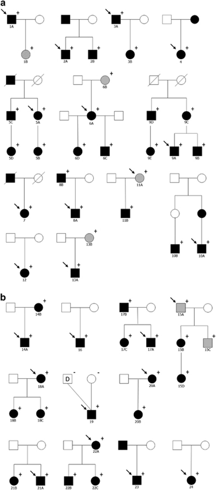
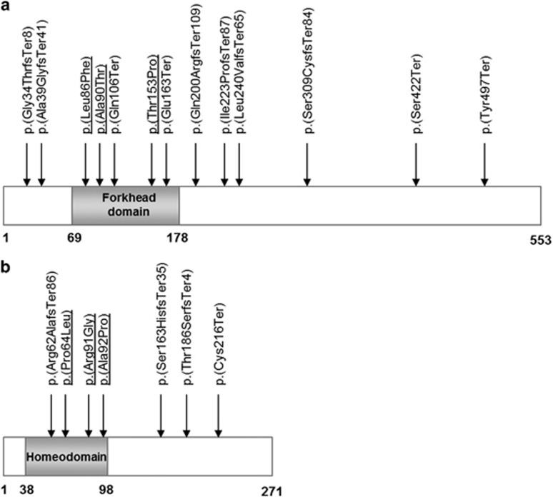
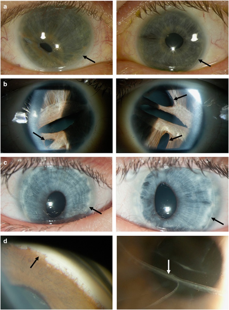
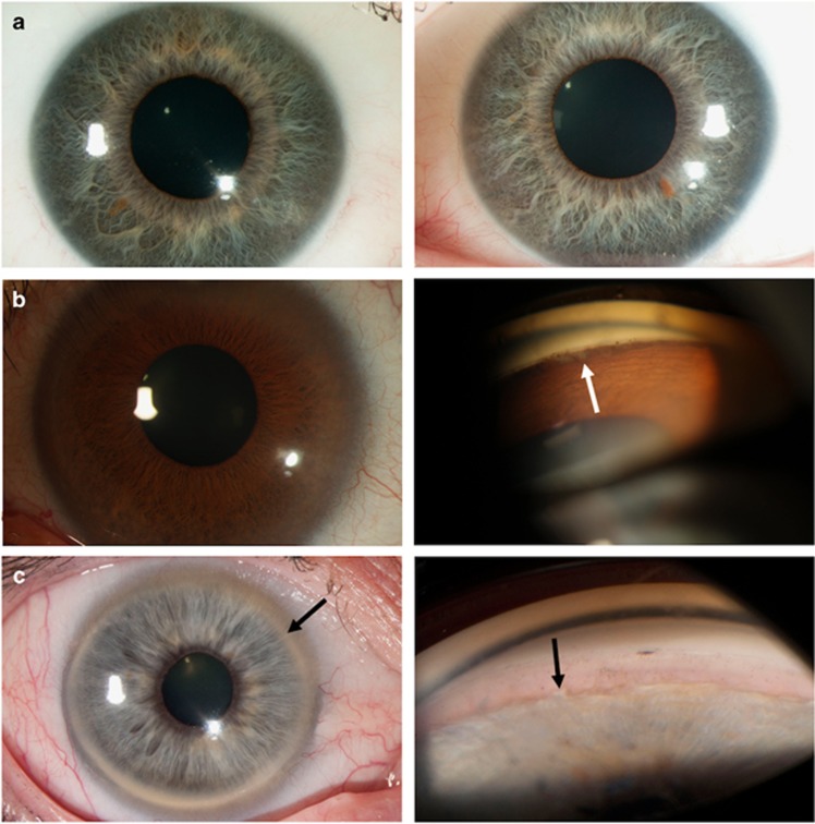
Variation in FOXC1 and PITX2 is associated with Axenfeld-Rieger syndrome, characterised by structural defects of the anterior chamber of the eye and a range of systemic features. Approximately half of all affected individuals will develop glaucoma, but the age at diagnosis and the phenotypic spectrum have not been well defined. As phenotypic heterogeneity is common, we aimed to delineate the age-related penetrance and the full phenotypic spectrum of glaucoma in FOXC1 or PITX2 carriers recruited through a national disease registry. All coding exons of FOXC1 and PITX2 were directly sequenced and multiplex ligation-dependent probe amplification was performed to detect copy number variation. The cohort included 53 individuals from 24 families with disease-associated FOXC1 or PITX2 variants, including one individual diagnosed with primary congenital glaucoma and five with primary open-angle glaucoma. The overall prevalence of glaucoma was 58.5% and was similar for both genes (53.3% for FOXC1 vs 60.9% for PITX2, P=0.59), however, the median age at glaucoma diagnosis was significantly lower in FOXC1 (6.0±13.0 years) compared with PITX2 carriers (18.0±10.6 years, P=0.04). The penetrance at 10 years old was significantly lower in PITX2 than FOXC1 carriers (13.0% vs 42.9%, P=0.03) but became comparable at 25 years old (71.4% vs 57.7%, P=0.38). These findings have important implications for the genetic counselling of families affected by Axenfeld-Rieger syndrome, and also suggest that FOXC1 and PITX2 contribute to the genetic architecture of primary glaucoma subtypes.
Conflict of interest statement
The authors declare no conflict of interest.
Figures
References
-
- Idrees F, Vaideanu D, Fraser SG, Sowden JC, Khaw PT: A review of anterior segment dysgeneses. Surv Ophthalmol 2006; 51: 213–231. - PubMed
-
- Shields MB, Buckley E, Klintworth GK, Thresher R: Axenfeld-Rieger syndrome. A spectrum of developmental disorders. Surv Ophthalmol 1985; 29: 387–409. - PubMed
-
- Alward WL: Axenfeld-Rieger syndrome in the age of molecular genetics. Am J Ophthalmol 2000; 130: 107–115. - PubMed
Publication types
MeSH terms
Substances
LinkOut - more resources
Full Text Sources
Other Literature Sources
Medical
Research Materials
