. 2017 Jun 18;16(12):1145-1152.
doi: 10.1080/15384101.2017.1320628. Epub 2017 May 17.

APC/C-Cdc20 mediates deprotection of centromeric cohesin at meiosis II in yeast

Affiliations

APC/C-Cdc20 mediates deprotection of centromeric cohesin at meiosis II in yeast

Katarzyna Jonak et al. Cell Cycle. .

Abstract

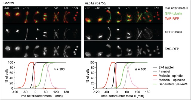
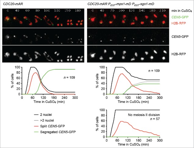
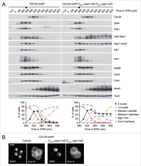
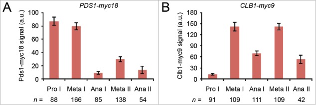
Cells undergoing meiosis produce haploid gametes through one round of DNA replication followed by 2 rounds of chromosome segregation. This requires that cohesin complexes, which establish sister chromatid cohesion during S phase, are removed in a stepwise manner. At meiosis I, the separase protease triggers the segregation of homologous chromosomes by cleaving cohesin's Rec8 subunit on chromosome arms. Cohesin persists at centromeres because the PP2A phosphatase, recruited by the shugoshin protein, dephosphorylates Rec8 and thereby protects it from cleavage. While chromatids disjoin upon cleavage of centromeric Rec8 at meiosis II, it was unclear how and when centromeric Rec8 is liberated from its protector PP2A. One proposal is that bipolar spindle forces separate PP2A from Rec8 as cells enter metaphase II. We show here that sister centromere biorientation is not sufficient to "deprotect" Rec8 at meiosis II in yeast. Instead, our data suggest that the ubiquitin-ligase APC/CCdc20 removes PP2A from centromeres by targeting for degradation the shugoshin Sgo1 and the kinase Mps1. This implies that Rec8 remains protected until entry into anaphase II when it is phosphorylated concurrently with the activation of separase. Here, we provide further support for this model and speculate on its relevance to mammalian oocytes.

Keywords: APC/C-Cdc20; Meiosis II; Mps1; PP2A; Rec8; centromere; cohesin cleavage; protection of centromeric cohesion; separase; shugoshin.


Figures

Figure 1.
Figure 1.
SET-related histone chaperones are dispensable for meiotic chromosome segregation in budding yeast. Spindles (GFP-tubulin), nuclei, and chromosome V sister sequences labeled with tet repressor (TetR) fused to the RFP tdTomato were imaged every 10 min in control (z21937) and nap1Δ vps75Δ (z21931) cells induced to enter meiosis. TetR-RFP bound to tet operators at the ura3 locus (ura3-tetO) on one copy of chromosome V produces one dot in meiosis I and 2 dots in meiosis II; free TetR-RFP illuminates the nucleoplasm. Top, representative time-lapse series. The width of one frame is 5 μm. Bottom, scoring of meiosis I and –II spindles, first (2 or 4 nuclei) and second (4 nuclei) division, and separated ura3-tetO sister sequences. Meiotic events were synchronised in silico by setting the time of meiosis II spindle formation (onset of metaphase II) to t = 0 in each cell.
Figure 2.
Figure 2.
Sister centromeres come under tension but fail to disjoin upon co-expression of non-degradable versions of Sgo1 and Mps1 in meiosis II. Control (z26823) and PEST-sgo1-mD PEST-mps1-mD (z26825) cells containing the CDC20-mAR synchronisation system were released from the metaphase I arrest with CuSO4 (10 μM, t = 0). Estradiol (10 μM) was added at t = 15 min to induce PEST-sgo1-mD and PEST-mps1-mD. Nuclei containing histone H2B tagged with the RFP mCherry and the sister centromeres of one chromosome V copy labeled with the CEN5-tetO/TetR-GFP system (CEN5-GFP) were imaged every 10 min. Top, representative time-lapse series. The width of one frame is 5 μm. Middle, percentages of cells with one division (2 nuclei), 2 divisions (more than 2 nuclei), split GFP signals (2 GFP-dots in the same nucleus), and segregated GFP signals (GFP signals in separate nuclei). Bottom right, PEST-sgo1-mD PEST-mps1-mD cells that fail to undergo the meiosis II division are plotted separately.
Figure 3.
Figure 3.
Co-expression of non-degradable Sgo1 and Mps1 in meiosis II does not interfere with silencing of the SAC. (A and B) Control (z26045) and PEST-sgo1-mD PEST-mps1-mD (z26015) cells containing the CDC20-mAR system were transferred to sporulation medium (SPM, t = 0) and released from the metaphase I arrest with CuSO4 at t = 480 min (arrow heads). Estradiol was added at t = 495 min. Note that endogenous and non-degradable Sgo1 carry Myc9 tags while both versions of Mps1 are tagged with Ha3. (A) Top, immunoblot detection of proteins in whole-cell extracts. Cyc, samples from proliferating cells. Bottom, percentages of cells with one (2 nuclei) or 2 divisions (more than 2 nuclei), meiosis I or meiosis II spindles, and spheres stained by FITC-concanavalin A (Con A spheres) were determined by immunofluorescence microscopy of fixed cells. Sgo1 foci were detected on chromatin spreads. 100 cells were counted per time point. (B) Cells stained with FITC-concanavalin A and DAPI at t = 640 min. Spheres around the nuclei represent mannoproteins in the prospore membrane and the first (inner) spore wall layer. Scale bar = 2 μm.
Figure 4.
Figure 4.
Levels of nuclear Pds1 and Clb1 at different stages of meiosis. (A and B) Formaldehyde-fixed cells from conventional meiotic cultures of untagged (z29971), PDS1-myc18 (z19647), and CLB1-myc9 (z29974) strains were stained with DAPI and antibodies to tubulin and Myc. The nuclear background signal generated by the α-Myc antibody was measured in 50–100 untagged cells at the indicated stage of meiosis, averaged, and the mean subtracted from the nuclear Myc signals of individual PDS1-myc18 (A) and CLB1-myc9 (B) cells at the corresponding meiotic stage. Columns indicate mean signal intensity in arbitrary units. Error bars show 95% confidence interval.
Figure 5.
Figure 5.
Levels and stability of separase/Esp1 in meiosis. (A and B) ESP1-myc18 strains were transferred to sporulation medium (SPM, t = 0) and treated with solvent (DMSO, 0.5%) or cycloheximide (CHX, 0.5 mg/ml) at the indicated times. Proteins were detected in whole-cell extracts by immunoblotting. Graphs show half-lives of Esp1-myc18 and Ama1 measured by scanning ECL signals on X-ray films. Data points are mean values from 2 gels. (A) Cells arresting at metaphase I due to CDC20 expression from the mitosis-specific CLB2 promoter (z30976) were treated with DMSO or CHX at t = 480 min. (B) CDC20-mAR cells (z29418) were released from the metaphase I arrest with CuSO4 at t = 480 min (arrowheads) and treated with DMSO or CHX at anaphase I (t = 520 min). Note that Dbf4 is degraded at anaphase I, Sgo1 at anaphase II, and Cdc5 at exit from meiosis II, whereas Ama1 is a constitutively unstable protein.

References

    1. Nasmyth K. Cohesin: a catenase with separate entry and exit gates? Nat Cell Biol 2011; 13:1170-7; PMID:21968990; https://doi.org/10.1038/ncb2349 - DOI - PubMed
    1. Haarhuis JHI, Elbatsh AMO, Rowland BD. Cohesin and Its Regulation: On the Logic of X-Shaped Chromosomes. Developmental Cell 2014; 31:7-18; PMID:25313959; https://doi.org/10.1016/j.devcel.2014.09.010 - DOI - PubMed
    1. Gligoris T, Lowe J. Structural Insights into Ring Formation of Cohesin and Related Smc Complexes. Trends Cell Biol 2016; 26:680-93; PMID:27134029; https://doi.org/10.1016/j.tcb.2016.04.002 - DOI - PMC - PubMed
    1. He X, Asthana S, Sorger PK. Transient sister chromatid separation and elastic deformation of chromosomes during mitosis in budding yeast. Cell 2000; 101:763-75; PMID:10892747; https://doi.org/10.1016/S0092-8674(00)80888-0 - DOI - PubMed
    1. Musacchio A. The Molecular Biology of Spindle Assembly Checkpoint Signaling Dynamics. Curr Biol 2015; 25:R1002-18. - PubMed

MeSH terms

LinkOut - more resources