. 2017 Dec;33(12):1071-1080.
doi: 10.1097/AJP.0000000000000509.

PPARγ Agonists Attenuate Trigeminal Neuropathic Pain

Affiliations

PPARγ Agonists Attenuate Trigeminal Neuropathic Pain

Danielle N Lyons et al. Clin J Pain. 2017 Dec.

Abstract

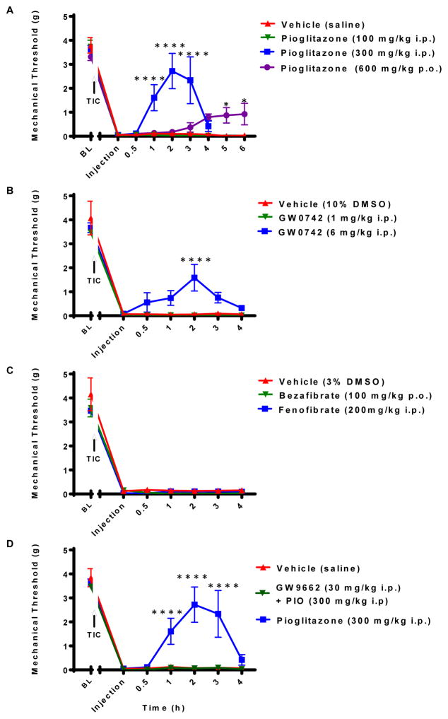
Objectives: The aim of this study is to investigate the role of peroxisome proliferator-activated receptor-gamma isoform (PPARγ), in trigeminal neuropathic pain utilizing a novel mouse trigeminal inflammatory compression (TIC) injury model.

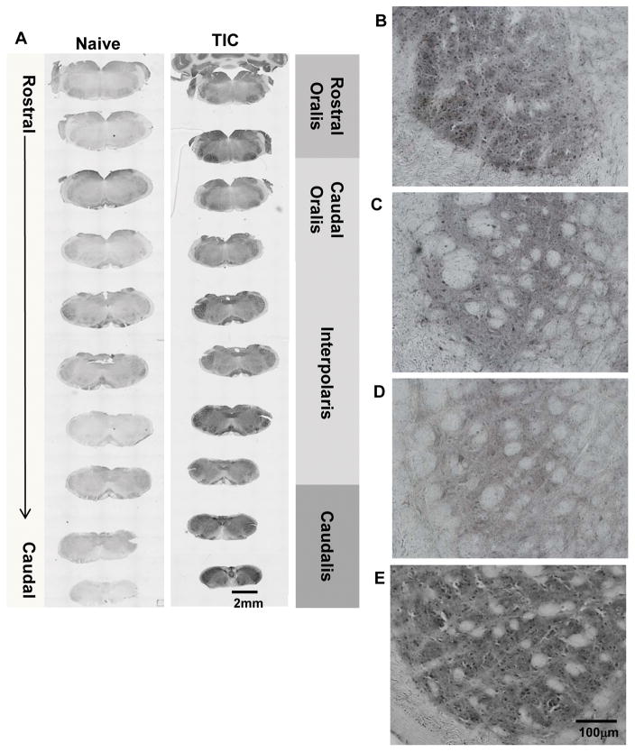
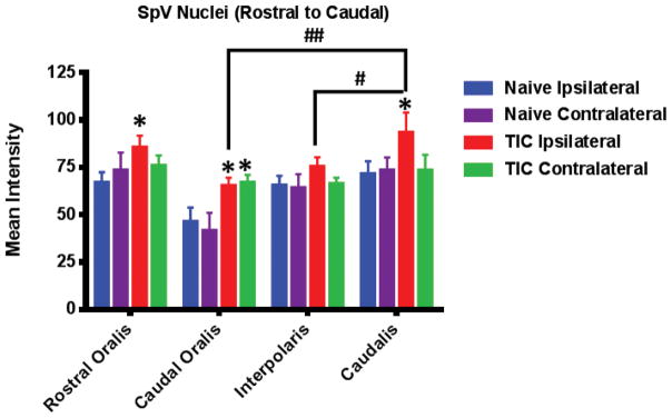
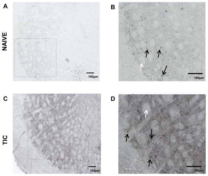
Results: The study determined that the PPARγ nuclear receptor plays a significant role in trigeminal nociception transmission, evidenced by: 1) Intense PPARγ immunoreactivity is expressed 3 weeks after TIC nerve injury in the spinal trigeminal caudalis, the termination site of trigeminal nociceptive nerve fibers. 2) Systemic administration of a PPARγ agonist, pioglitazone (PIO), attenuates whisker pad mechanical allodynia at doses of 300 mg/kg i.p. and 600 mg/kg p.o. 3) Administration of a PPARγ antagonist, GW9662 (30 mg/kg i.p.), prior to providing the optimal dose of PIO (300 mg/kg i.p.) blocked the analgesic effect of PIO.

Discussion: This is the first study localizing PPARγ immunoreactivity throughout the brainstem trigeminal sensory spinal nucleus (spV) and its increase three weeks after TIC nerve injury. This is also the first study to demonstrate that activation of PPARγ attenuates trigeminal hypersensitivity in the mouse TIC nerve injury model. The findings presented here suggest the possibility of utilizing the FDA approved diabetic treatment drug, PIO, as a new therapeutic that targets PPARγ for treatment of patients suffering from orofacial neuropathic pain.


Figures

Figure 1
Figure 1. Mice with TIC Injury Developed Persistent Unilateral Mechanical Allodynia on the Ipsilateral Whisker Pad
The 50% mechanical threshold on whisker pads of the mice with TIC injury and the sham mice were measured bilaterally for detecting mechanical allodynia. The mechanical threshold on the ipsilateral whisker pad of mice with TIC injury was significantly decreased within one week of injury and remained decreased until euthanasia, week 14 (indicated with thin black line). The mechanical threshold on contralateral whisker pad of the mice with TIC injury was unaffected by the surgery. The mechanical threshold on the whisker pads of the sham operation mice did not change. (n=8/group; ****p<0.0001, two-way ANOVA, Fisher’s post hoc test)
Figure 2
Figure 2. PIO Attenuated Whisker Pad Mechanical Allodynia in the Mice with TIC Injury
Hypersensitivity was attenuated by specific PPARγ agonism with (A) PIO rapidly elevating the 50% mechanical threshold in the mice with TIC at higher doses (300 mg/kg and 600 mg/kg), but was ineffective at 100 mg/kg (n=3–7). (B) GW0742, PPARβ agonist, attenuated mechanical allodynia in the mice with TIC injury at a dose of 6 mg/kg, but was not as effective as PIO (n=4–6). (C) PPARα agonists, Bezafibrate and fenofibrate at indicated doses, were not effective in alleviating mechanical allodynia in the mice with TIC injury (n=3–6). (D) PPARγ antagonist, GW9662, blocked the anti-allodynic effect of PIO at a dose of 30 mg/kg (n=4–7). (*p<0.05, ****p<0.0001; two-way ANOVA, Fisher’s post hoc test)
Figure 3
Figure 3. PPARγ Localization Throughout SpV of Mice with TIC Injury
(A) The photomicrographic images depict the rostral to caudal distribution of PPARγ in the spV nucleus of naïve and TIC injured mice. PPARγ is localized throughout most of the spV in TIC injured mice and is more abundant than in naïve mice. The low power images show intensity differences in PPARγ among individual spV subnuclei (rostral and caudal oralis, interpolaris, caudalis). Density differences are seen in the rostral (B) versus caudal (C) oralis subnuclei in mice with TIC injury. (D) Less density for PPARγ is localized in spV subnucleus interpolaris in mice with TIC injury. (E) Dense localization of PPARγ is evident in spV caudalis subnucleus in mice with TIC injury.
Figure 4
Figure 4. PPARγ Immunoreactivity Increased in spV of Mice with TIC Injury
The mice with TIC injury had an increased PPARγ immunoreactivity in the ipsilateral spV rostral oralis and subnucleus caudalis compared to naïve mice (n=3 with 9–12 sections/animal (*p<0.05, two-way ANOVA, Fisher’s post hoc). Mean intensity of immunofluorescence is shown along the Y-axis. The spV caudal oralis subnucleus expressed bilateral immunoreactivity that was significant greater than tissue from naïve mice (*p<0.05, two-way ANOVA, Fisher’s post hoc). PPARγ immunoreactivity was also significantly higher in ipsilateral spV caudalis in mice with TIC injury compared spV oralis and interpolaris. (#p<0.05, ##p<0.01, two-way ANOVA, Fisher’s post hoc)
Figure 5
Figure 5. PPARγ Immunoreactivity in SpV Interpolaris in Sham and TIC Injured Mice
The upper panels illustrate PPARγ in ipsilateral spV dorsal horn where most PPARγ positive cells were most apparent shown from sham mice at lower (A) and higher magnification (B). The lower panels (C) and (D) illustrate ipsilateral side PPARγ in mice with TIC injury. The black arrows indicate PPARγ positive cells and the white arrows indicate the whisker barrel axonal projections from the mouse whisker pad receptive fields. As shown by the pictures, the staining intensity of PPARγ immunoreactivity was more intense in the tissue sections from the TIC injured mice than from the sham mice.

Similar articles

Cited by

References

    1. Zakrzewska JM. Differential diagnosis of facial pain and guidelines for management. Br J Anaesth. 2013;111:95–104. - PubMed
    1. Asmundson GJ, Katz J. Understanding the co-occurrence of anxiety disorders and chronic pain: state-of-the-art. Depress Anxiety. 2009;26:888–901. - PubMed
    1. De Leeuw R. Orofacial Pain: Guidelines for Assessment, Diagnosis, and Management. 4. Hanover Park, IL: Quintessence Publishing Co, Inc; 2008. p. 316.
    1. Robinson MJ, Edwards SE, Iyengar S, Bymaster F, Clark M, Katon W. Depression and pain. Front Biosci (Landmark Ed) 2009;14:5031–51. - PubMed
    1. Roditi D, Robinson ME, Litwins N. Effects of coping statements on experimental pain in chronic pain patients. J Pain Res. 2009;2:109–16. - PMC - PubMed

MeSH terms