

Infected tibia defect fractures treated with the Masquelet technique
- PMID: 28514314
- PMCID: PMC5440151
- DOI: 10.1097/MD.0000000000006948
Infected tibia defect fractures treated with the Masquelet technique
Abstract
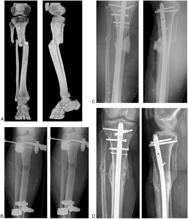
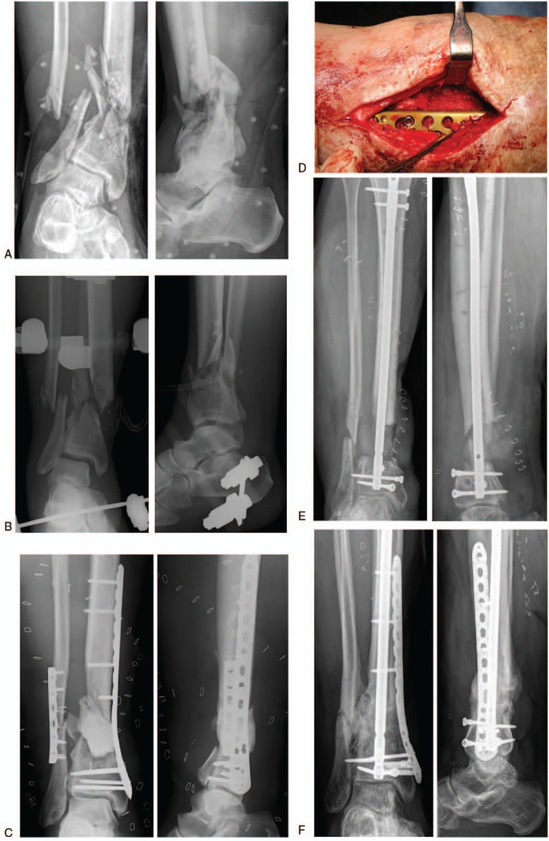
The treatment after open and infected fractures with extensive soft tissue damage and bone deficit remains a challenging clinical problem. The technique described by Masquelet, using a temporary cement spacer to induce a membrane combined with reconstructive soft tissue coverage, is a possible solution. This study describes the work-up, operative procedure, complications, and the outcome of a homogenous group of patients with an open and infected tibia fracture and segmental bone loss treated with the Masquelet technique (MT).This retrospective study evaluates patients having sustained an open tibia fracture treated with the MT between 2008 and 2013 with a follow up of at least 1 year. The defect was either primary, caused by a high-grade open fracture or secondary due to a non-union after an open fracture. Prerequisite conditions prior to the procedure of the Masquelet were a defect zone with eradicated infection, an intact soft tissue cover and stability provided by an external fixation.Volume of the defect, time until the implantation of the spacer, time of the spacer in situ and the time to clinical and radiological union were evaluated. Patient records were screened for reoperations and complications. The functional clinical outcome was measured.Eight patients were treated with a follow up over 1 year. The spacer was implanted after a median of 11 (2-70) weeks after the accident. The predefined conditions for the Masquelet phase were reached after a median of 12 (7-34) operations.Seven patients required reconstructive soft tissue coverage. The volume of the defect had a median of 111 (53.9-621.6) cm, the spacer was in situ for a median of 12 (7-26) weeks. Radiological healing was achieved in 7 cases after a median time of 52 (26-93) weeks.Full weight bearing was achieved after a median time of 16 (11-24) weeks. Four patients needed a reoperation. The lower limb functional index was a median of 60% (32-92%).Seven out of 8 patients treated in this group of severe open and infected tibia fractures did both clinically and radiologically heal. Due to the massive destruction of the soft tissue, patients needed several reoperations with soft tissue debridements and reconstruction before the spacer and the bone graft could be implanted.
Conflict of interest statement
The authors report no conflicts of interest.
Figures
Similar articles
-
The Masquelet technique in the treatment of a non-infected open complex fracture of the distal tibia with severe bone and soft tissue loss: A case report.Injury. 2018 Dec;49 Suppl 4:S58-S62. doi: 10.1016/j.injury.2018.11.039. Epub 2018 Dec 3. Injury. 2018. PMID: 30526950
-
[Muscle flap transfer of the treatment of infected tibial and malleolar fractures and chronic osteomyelitis of the tibia].Acta Chir Orthop Traumatol Cech. 2007 Jun;74(3):162-70. Acta Chir Orthop Traumatol Cech. 2007. PMID: 17623603 Czech.
-
Bifocal compression-distraction in the acute treatment of grade III open tibia fractures with bone and soft-tissue loss: a report of 24 cases.J Orthop Trauma. 2004 Mar;18(3):150-7. doi: 10.1097/00005131-200403000-00005. J Orthop Trauma. 2004. PMID: 15091269
-
"Primary free-flap tibial open fracture reconstruction with the Masquelet technique" and internal fixation.Injury. 2020 Dec;51(12):2970-2974. doi: 10.1016/j.injury.2020.10.039. Epub 2020 Oct 8. Injury. 2020. PMID: 33097199 Review.
-
[Reconstruction of osseous defects using the Masquelet technique].Orthopade. 2017 Aug;46(8):665-672. doi: 10.1007/s00132-017-3443-1. Orthopade. 2017. PMID: 28744608 Review. German.
Cited by
-
The Concept of Scaffold-Guided Bone Regeneration for the Treatment of Long Bone Defects: Current Clinical Application and Future Perspective.J Funct Biomater. 2023 Jun 27;14(7):341. doi: 10.3390/jfb14070341. J Funct Biomater. 2023. PMID: 37504836 Free PMC article. Review.
-
Functional outcomes and health-related quality of life after reconstruction of segmental bone loss in femur and tibia using the induced membrane technique.Arch Orthop Trauma Surg. 2023 Aug;143(8):4587-4596. doi: 10.1007/s00402-022-04714-9. Epub 2022 Dec 3. Arch Orthop Trauma Surg. 2023. PMID: 36460763 Free PMC article.
-
Use of locked plates and mono-rail fixator in segmental tibial defects: A prospective interventional study.J Orthop. 2023 Aug 14;44:47-52. doi: 10.1016/j.jor.2023.08.003. eCollection 2023 Oct. J Orthop. 2023. PMID: 37664557 Free PMC article.
-
Eradication of Acinetobacter baumannii/Enterobacter cloacae complex in an open proximal tibial fracture and closed drop foot correction with a multidisciplinary approach using the Taylor Spatial Frame®: a case report.Eur J Med Res. 2019 Jan 19;24(1):2. doi: 10.1186/s40001-019-0360-2. Eur J Med Res. 2019. PMID: 30660181 Free PMC article.
-
Reconstruction of gap non-union tibia with composite use of extramedullary fixation and bone transport by monorail fixator: a prospective case series.J West Afr Coll Surg. 2024 Jul-Sep;14(3):324-330. doi: 10.4103/jwas.jwas_152_23. Epub 2024 May 24. J West Afr Coll Surg. 2024. PMID: 38988428 Free PMC article.
References
-
- Weiland AJ, Phillips TW, Randolph MA. Bone grafts: a radiologic, histologic, and biomechanical model comparing autografts, allografts, and free vascularized bone grafts. Plast Reconstr Surg 1984;74:368–79. - PubMed
-
- Masquelet AC, Fitoussi F, Begue T, et al. Reconstruction of the long bones by the induced membrane and spongy autograft. Ann Chir Plast Esthet 2000;45:346–53. - PubMed
-
- Aho O, Lehenkari P, Ristiniemi J, et al. The mechanism of action of induced membranes in bone repair. J Bone Joint Surg Am 2013;95:597–604. - PubMed
-
- Pelissier P, Masquelet AC, Bareille R, et al. Induced membranes secrete growth factors including vascular and osteoinductive factors and could stimulate bone regeneration. J Orthop Res 2004;22:73–9. - PubMed
Publication types
MeSH terms
LinkOut - more resources
Full Text Sources
Other Literature Sources
Medical
