. 2017 May 25;545(7655):482-486.
doi: 10.1038/nature22365. Epub 2017 May 17.

Evolutionary enhancement of Zika virus infectivity in Aedes aegypti mosquitoes

Affiliations

Evolutionary enhancement of Zika virus infectivity in Aedes aegypti mosquitoes

Yang Liu et al. Nature. .

Abstract

Zika virus (ZIKV) remained obscure until the recent explosive outbreaks in French Polynesia (2013-2014) and South America (2015-2016). Phylogenetic studies have shown that ZIKV has evolved into African and Asian lineages. The Asian lineage of ZIKV was responsible for the recent epidemics in the Americas. However, the underlying mechanisms through which ZIKV rapidly and explosively spread from Asia to the Americas are unclear. Non-structural protein 1 (NS1) facilitates flavivirus acquisition by mosquitoes from an infected mammalian host and subsequently enhances viral prevalence in mosquitoes. Here we show that NS1 antigenaemia determines ZIKV infectivity in its mosquito vector Aedes aegypti, which acquires ZIKV via a blood meal. Clinical isolates from the most recent outbreak in the Americas were much more infectious in mosquitoes than the FSS13025 strain, which was isolated in Cambodia in 2010. Further analyses showed that these epidemic strains have higher NS1 antigenaemia than the FSS13025 strain because of an alanine-to-valine amino acid substitution at residue 188 in NS1. ZIKV infectivity was enhanced by this amino acid substitution in the ZIKV FSS13025 strain in mosquitoes that acquired ZIKV from a viraemic C57BL/6 mouse deficient in type I and II interferon (IFN) receptors (AG6 mouse). Our results reveal that ZIKV evolved to acquire a spontaneous mutation in its NS1 protein, resulting in increased NS1 antigenaemia. Enhancement of NS1 antigenaemia in infected hosts promotes ZIKV infectivity and prevalence in mosquitoes, which could have facilitated transmission during recent ZIKV epidemics.


Conflict of interest statement

Author information

The authors declare that they have no competing financial interests.

Figures
Extended Data Figure 1
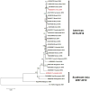
Extended Data Figure 1. Phylogenetic analysis of ZIKV isolates in the Asian lineage
The tree was constructed using the neighbor-joining (NJ) method based on the alignment of ZIKV sequences in the Asian lineage. The bootstrap values of 2000 replicates are indicated on the branch nodes.
Extended Data Figure 2
Extended Data Figure 2. Generation of murine anti-ZIKV NS1 polyclonal antibody and purification of the GZ01 ZIKV NS1 protein from Drosophila S2 cells
a, Generation of murine anti-ZIKV NS1 polyclonal antibody. A murine ZIKV NS1 polyclonal antibody was generated by immunization of the purified GZ01 ZIKV NS1 recombinant protein expressed in E. coli. The antibody was validated by probing an S2-expressed ZIKV NS1 recombinant protein. The same samples probed by murine pre-immune antibody served as a negative control. b, Purification of the GZ01 ZIKV NS1 protein from Drosophila S2 cells. Full-length NS1 was cloned into the pMT/BiP/V5-His A expression vector. Recombinant ZIKV NS1 protein was expressed in Drosophila S2 cells and purified using a cobalt-His column (Left Panel). Protein quality was evaluated by Western-blotting with an anti-V5 antibody (Right Panel). a, b, The experiments were reproduced 3 times with similar results. For gel source data, see Supplementary Fig. 1.
Extended Data Figure 3
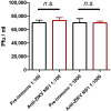
Extended Data Figure 3. Anti-ZIKV NS1 antibodies did not neutralize ZIKV virions
We premixed two dilutions of murine ZIKV NS1 polyclonal antisera (1:100 and 1:1000) with purified ZIKV GZ01 infectious virions. The same amount of pre-immune sera served as a negative control. After a 30 min incubation, the infectivity of the "antibody-ZIKV virion" mixture was determined by plaque assay on Vero cells. The values in the graph represent the mean ± s.e.m. p values were determined by two-tailed Mann-Whitney test. The data were combined from 3 biological repeats. n.s., not significant.
Extended Data Figure 4
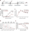
Extended Data Figure 4. Passive transfer of ZIKV NS1 antibodies into infected AG6 mice prevented ZIKV acquisition by A. aegypti
a, Schematic representation of the study design. AG6 mice were intradermally infected with 1×10⁴ pfu of the ZIKV GZ01 strain. Subsequently, 100 μl of a murine ZIKV NS1 antibody was intraperitoneally inoculated into the mice 12 hr post-infection. The same amount of pre-immune serum was inoculated as a mock control. After 12 hr of antibody dissemination, the infected mice were subjected to daily biting by female A. aegypti from day 1 to day 5 post-mouse infection. The mouse blood-fed mosquitoes were reared for an additional 8 days for ZIKV detection. b, ZIKV NS1 measurement by ELISA (n=6 mice per group pooled from 3 independent biological replicates). Mouse sera were collected to quantify the amounts of ZIKV NS1 protein from days 0 and 6 post-mouse infection. c, Detection of the ZIKV load in the blood of the infected mice (n=8 mice per group pooled from 4 independent biological replicates). The presence of infectious ZIKV particles in blood plasma was determined by a plaque assay from days 0 to 6 post-mouse infection. d, e, Immuno-blockade of ZIKV NS1 in infected AG6 mice reduced the infection of fed A. aegypti (n=6 mice per group pooled from 3 independent biological replicates). The number of infected mosquitoes relative to the total number of mosquitoes is shown at the top of each column. Each dot represents a mosquito (d). The data are represented as the percentage of mosquito infection (e). Data are mean ± s.e.m. (b, c). p values were determined by two-tailed Mann-Whitney test (b-d) or two-sided Fisher’s exact test (e). **p< 0.01, ***p< 0.001, n.s., not significant.
Extended Data Figure 5
Extended Data Figure 5. Modeling the location of the 188th residue of ZIKV NS1 and assessing NS1 dimer formation in cell lysates
a, Modeling the location of the 188th residue of ZIKV NS1. Modeling was based on the ZIKV NS1 structure (PDB: 5K6K). The 188th position is located within the interface of a NS1 dimer (contoured by red), which is formed by hydrophobic interactions. The 188th amino acid is deep within the hydrophobic pocket formed by Trp-210, Leu-23, His-299 and Leu-231. b, Assessing NS1 dimer formation in the lysates of cells transfected with GZ01 or FSS13025 prM-E-NS1 recombinant plasmids. The cell lysates, with or without heating treatment, were used for assessing NS1 monomer and dimer by Western blotting with an anti-V5 antibody, respectively. The experiment was reproduced 3 times with similar results. For gel source data, see Supplementary Fig. 1.
Extended Data Figure 6
Sequence alignment of NS1 sequences in ZIKV isolates of the Asian lineage.
Extended Data Figure 7
Extended Data Figure 7. A single amino acid substitution enhanced NS1 secretability and ZIKV infectivity in mosquitoes
a–c, NS1 secretability in ZIKV-infected Vero cells. We mutated the single amino acid from alanine to valine at the 188th position of NS1 protein on an infectious clone of ZIKV Cambodian FSS13025 strain (FSS13025-A188V strain). FSS13025 or FSS13025-A188V strain (0.01 M.O.I.) was used to infect Vero cells. Supernatants from the infected Vero cells were collected from 0 to 5 days post-infection. a, The amount of ZIKV NS1 protein was determined by ELISA. b, ZIKV titer was determined by a plaque assay. c, NS1 quantity normalized against average viral titer (ng per pfu). The data were pooled from 3 independent biological replicates. d–e, NS1 secretability determined ZIKV infectivity in mosquitoes. The supernatant, isolated from the FSS13025-infected or FSS13025-A188V-infected Vero cells, was premixed with fresh human blood at a 1:1 ratio for an in vitro membrane feeding experiment (final titer is 5×10⁴ pfu/ml), respectively. The fed mosquitoes were scarified on 8 days after a blood meal to determine the ZIKV load by qPCR. The number of infected mosquitoes relative to the total number of mosquitoes (infected number/total number) is shown at the top of each column. Each dot represents a mosquito (d). The data are represented as the percentage of mosquito infections (e). The data were pooled from 3 independent biological replicates. Data are mean ± s.e.m. (a-c). p values were determined by two-tailed Mann-Whitney test (a-d) or two-sided Fisher’s exact test (e). *p< 0.05, **p< 0.01, n.s., not significant.
Extended Data Figure 8
Extended Data Figure 8. Neutralization of NS1 by murine ZIKV NS1 antisera effectively reduced the prevalence of FSS13025-A188V ZIKV in A. aegypti
a, b, Infection by FSS13025-A188V ZIKV in AG6 mice. The AG6 mice were intradermally inoculated with these viruses (1×10⁴ pfu). ZIKV NS1 measurement by ELISA. Mouse sera were collected to qualify the amounts of ZIKV NS1 protein from days 0 to 6 post-mouse infection (a). Detection of the ZIKV load in the blood of infected AG6 mice. The presence of infectious ZIKV particles in blood plasma was determined by a plaque assay (b). n=10 mice per group pooled from 4 independent biological replicates. c, d, Immuno-blockade of NS1 in the FSS13025-A188V ZIKV infected AG6 mice reduced the acquisition of fed A. aegypti (n=7 mice per group pooled from 3 independent biological replicates). The number of infected mosquitoes relative to the total number of mosquitoes is shown at the top of each column. Each dot represents a mosquito (c). The data are represented as the percentage of mosquito infection (d). Data are mean ± s.e.m. (a, b). p values were determined by two-tailed Mann-Whitney test (a-c) or two-sided Fisher’s exact test (d). *p< 0.05, ***p< 0.001, n.s., not significant.
Extended Data Figure 9
Extended Data Figure 9. Sequence alignment of NS1 sequences in African ZIKV isolates, and NS1 secretability in African ZIKV-infected Vero cells
a, Sequence alignment of NS1 sequences in African ZIKV isolates. b-d, NS1 secretability in African ZIKV-infected Vero cells. Vero cells were infected at an M.O.I. of 0.01 with the GZ01, FSS13025 and MR766 strains. Supernatants from the infected Vero cells were collected from 0 to 4 days post-infection. b, ZIKV titer was determined by a plaque assay. c, The amount of ZIKV NS1 protein was determined by ELISA. d, NS1 quantity normalized against average viral titer (ng per pfu). The data were pooled from 3 independent biological replicates. The values in the graph represent the mean ± s.e.m. (b-d).
Figure 1
Figure 1. Comparing the infectivity of ZIKV isolates of Asian lineage in mosquitoes
a, Schematic representation of the study design. Separated groups of AG6 mice were intradermally infected with 1×10⁴ pfu of the GZ01 and FSS13025 strains. b, Detection of the ZIKV load in the blood plasma by a plaque assay (n=14 mice per group pooled from 5 independent biological replicates). c, ZIKV NS1 measurement by ELISA (n=6 mice per group pooled from 3 independent biological replicates). d, e, Comparison of the infectivity of two ZIKV isolates in A. aegypti (n=6 mice per group pooled from 3 independent biological replicates). The number at the top of each column represents infected number/total number. Each dot represents a mosquito (d). The data are represented as the percentage of mosquito infections (e). Data are mean ± s.e.m. (b, c). p values were determined by two-tailed Mann-Whitney test (b-d) or two-sided Fisher’s exact test (e). **p< 0.01, ***p< 0.001, n.s., not significant.
Figure 2
Figure 2. Differences in NS1 secretion determine ZIKV infectivity in mosquitoes
a-c, NS1 secretability in ZIKV-infected Vero cells. Vero cells were infected with 0.01 M.O.I. viruses. c, NS1 quantity normalized against average viral titer (ng per pfu). The data were pooled from 3 independent biological replicates. d-f, Immunoblockade of NS1 in GZ01-infected supernatants reduced ZIKV acquisition. Either 10 μl of murine ZIKV NS1 antisera or pre-immune sera were mixed with supernatant from ZIKV GZ01-infected Vero cells (500 μl) and fresh human blood (500 μl) for in vitro membrane feeding of A. aegypti. The data were pooled from 2 independent biological replicates. g-i, Addition of NS1 in FSS13025-infected supernatant increased the mosquito infection. Either 10 μg of purified NS1 or BSA was incubated with fresh human blood (500 μl) and supernatant from ZIKV FSS13025-infected Vero cells (500 μl) to feed A. aegypti. The data were pooled from 2 independent biological replicates. j-l, Assessment of the threshold NS1 concentration that enhances ZIKV acquisition. Serial dilutions of purified ZIKV NS1 (10 μl) were mixed with purified GZ01 ZIKV virions (10 μl) and fresh human blood (1000 μl) for A. aegypti feeding. The data were pooled from 4 independent biological replicates. A final concentration of 1×10⁵ pfu/ml ZIKV was used for mosquito oral infection (d, g, j). The number at the top of each column represents infected number/total number. Each dot represents a mosquito (e, h, k). Data are mean ± s.e.m. (a-c). p values were determined by two-tailed Mann-Whitney test (e, h, k) or two-sided Fisher’s exact test (f, i, l), and adjusted using Bonferroni correction to account for multiple comparisons (k, l). The p value represents a comparison between BSA and other groups (k, l). *p< 0.0125, ***p< 0.00025, n.s., not significant.
Figure 3
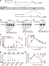
Figure 3. A single amino acid residue in NS1 determines its secretability in the Asian lineage of ZIKV
a, Comparison of the prM-E-NS1 sequence between GZ01 and FSS13025 isolates. b, Detection of NS1 secretion in ectopically expressed prM-E-NS1 proteins of the subcloned and mutated GZ01 and FSS13025 strains. c, Detection of NS1 secretion in ectopically expressed prM-E-NS1 proteins of ZIKV Asian strains. d, Mutation of alanine to valine in the NS1 protein conferred NS1 secretability to the prM-E-NS1 proteins of a Malaysia P6-740 strain. b-d, The experiments were reproduced 3 times with similar results. For gel source data, see Supplementary Fig. 1. e, Measurement of ZIKV NS1 antigenemia by ELISA (n=10 mice per group pooled from 5 independent biological replicates). f, Detection of the ZIKV load in the mouse blood by a plaque assay (n=8 mice per group pooled from 4 independent biological replicates). g, h, Comparison of mosquito infectivity (g) and infective ratio (h) of ZIKV strains (n=6 mice per group pooled from 3 independent biological replicates). The number at the top of each column represents infected number/total number. Each dot represents a mosquito (g). The AG6 mice were intradermally inoculated with 1×10⁴ pfu of the FSS13025 and FSS13025-A188V strains, data are mean ± s.e.m. (e, f). p values were determined by two-tailed Mann-Whitney test (e-g) or two-sided Fisher’s exact test (h). *p< 0.05, ***p< 0.001, n.s., not significant.
Figure 4
Figure 4. A single amino acid substitution influences the prevalence of mosquito infection during the "mosquito-host-mosquito" life cycle
a, Schematic representation of the study design. b, Detection of the ZIKV viremia by a plaque assay (n=12 mice per group pooled from 4 independent biological replicates). c, Measurements of ZIKV NS1 antigenemia by ELISA (n=12 mice per group pooled from 4 independent biological replicates). d, e, Comparison of mosquito infectivity (d) and infective ratio (e) of ZIKV strains (n=9 mice per group pooled from 3 independent biological replicates). The number at the top of each column represents infected number/total number. Each dot represents a mosquito (d). Data are mean ± s.e.m. (b, c). p values were determined by two-tailed Mann-Whitney test (b-d) or two-sided Fisher’s exact test (e), and adjusted using Bonferroni correction to account for multiple comparisons (b-e). The black p value represents a comparison between GZ01 and FSS13025. The blue p value represents a comparison between FSS13025-A188V and FSS13025. *p< 0.025, **p< 0.005, ***p< 0.0005, n.s., not significant.

Comment in

References

    1. Marchette NJ, Garcia R, Rudnick A. Isolation of Zika virus from Aedes aegypti mosquitoes in Malaysia. Am J Trop Med Hyg. 1969;18:411–415. - PubMed
    1. Musso D, Gubler DJ. Zika Virus. Clin Microbiol Rev. 2016;29:487–524. - PMC - PubMed
    1. Oehler E, et al. Zika virus infection complicated by Guillain-Barre syndrome-case report, French Polynesia, December 2013. Eurosurveillance. 2014;19:4–6. - PubMed
    1. Cordeiro MT, Pena LJ, Brito CA, Gil LH, Marques ET. Positive IgM for Zika virus in the cerebrospinal fluid of 30 neonates with microcephaly in Brazil. Lancet. 2016;387:1811–1812. - PubMed
    1. Faye O, et al. Molecular evolution of Zika virus during its emergence in the 20th century. Plos Neglect Trop D. 2014;8:2–3. - PMC - PubMed

Publication types

MeSH terms

Substances