. 2017 May 16;46(5):792-803.e3.
doi: 10.1016/j.immuni.2017.04.014.

Glycine Substitution at Helix-to-Coil Transitions Facilitates the Structural Determination of a Stabilized Subtype C HIV Envelope Glycoprotein

Affiliations

Glycine Substitution at Helix-to-Coil Transitions Facilitates the Structural Determination of a Stabilized Subtype C HIV Envelope Glycoprotein

Javier Guenaga et al. Immunity. .

Abstract

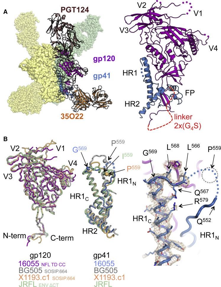
Advances in HIV-1 envelope glycoprotein (Env) design generate native-like trimers and high-resolution clade A, B, and G structures and elicit neutralizing antibodies. However, a high-resolution clade C structure is critical, as this subtype accounts for the majority of HIV infections worldwide, but well-ordered clade C Env trimers are more challenging to produce due to their instability. Based on targeted glycine substitutions in the Env fusion machinery, we defined a general approach that disfavors helical transitions leading to post-fusion conformations, thereby favoring the pre-fusion state. We generated a stabilized, soluble clade C Env (16055 NFL) and determined its crystal structure at 3.9 Å. Its overall conformation is similar to SOSIP.664 and native Env trimers but includes a covalent linker between gp120 and gp41, an engineered 201-433 disulfide bond, and density corresponding to 22 N-glycans. Env-structure-guided design strategies resulted in multiple homogeneous cross-clade immunogens with the potential to advance HIV vaccine development.

Keywords: Antibody; Envelope glycoprotein; Glycan shield; HIV; Immunogen; Trimer; Vaccine; bNAb.


Figures

None
Graphical abstract
Figure 1
Figure 1
Env gp41 Glycine Substitutions Increase Native-like Trimer Formation (A) Schematic model of the targeted glycine strategy to attenuate helical secondary structural transitions to the post-fusion form (left) and location of the tested glycine substitutions (red spheres) displayed on the crystal structure of the gp41 subunit in BG505 SOSIP.664 (PDB: 5CEZ) (right). (B) SEC profiles of subtype C 1086c NFL TD variants with glycine substitutions at the indicated residues and TD as a control with no glycines. The vertical dotted red line denotes the elution volume of native-like, well-ordered trimers. (C) SEC profiles of HIV-1 subtype C 16055 NFL TD trimers with and without T569G after lectin chromatography purification. Shown in the upper right is the SEC profile corresponding to a re-run of the trimer fractions. 2D class averages from negative-stain EM of the trimer fraction is shown below. See also Figures S1 and S2.
Figure 2
Figure 2
The Cleavage-Independent Subtype C HIV-1 Env 16055 NFL TD CC Trimer Shares Homology with Other Env Trimer Structures (A) Crystal structure at 3.9 Å resolution of the 16055 NFL TD CC (T569G) trimer (gp120 purple, gp41 blue, PDB: 5UM8) in complex with Fabs PGT124 (brown) and 35O22 (orange). (B) Superimposition of the gp120 subunits of soluble Env trimers derived from HIV-1 clade C NFL (magenta), clade A BG505 SOSIP.664 (gray, PDB: 5CEZ), clade G X1193.c1 SOSIP.664 (light orange, PDB: 5FYJ), and clade B JRFL Env ΔCT (green, PDB: 5FUU) (left). Corresponding overlap of the gp41 subunits of 16055 (blue), BG505 (gray), X1193.c1 (light orange), and JRFL Env (green) (middle). Close-up view of the HR1 region of the NFL trimer showing the T569G mutation and residues downstream that demarcate the region that undergoes the coil-to-helix transition in the post-fusion structure (right). See also Figures S2 and S3.
Figure 3
Figure 3
N-Glycosylation Sites in the 16055 NFL TD CC Structure Differ from Those in the BG505 SOSIP.664 Structure (A) Schematic representation of the 16055 NFL and BG505 SOSIP trimer constructs, with all PNGS numbered on Env regions and those visible in either structure highlighted in green boxes. (B) 16055 NFL TD CC (T569G) structure (top) with gp120 (magenta), gp41 (blue), and BG505 SOSIP.664 structure (where N137 was deleted) (bottom, gray) (PDB: 5CEZ) in cartoon representation, with visible N-glycans as green or blue spheres. Blue spheres represent glycans visible in the 16055 NFL TD CC (T569G) structure that are either not ordered in the BG505 SOSIP.664 structure or are not PNGS in BG505 Env, and the orange spheres represent the reverse in BG505 SOSIP.664.
Figure 4
Figure 4
PGT124 Displays Broad Recognition of the Glycan Shield, while Autologous NAbs Exploit Gaps in the Shield (A) 16055 NFL TD CC (T569G) structure with gp120 (magenta), gp41 (blue) (left), and BG505 SOSIP.664 structure (gray, right) in cartoon and surface-mode representation, with glycans as green or yellow spheres. Yellow spheres denote glycans present on 16055 Env that are not present in BG505 Env that produce a glycan hole that is targeted by vaccine-elicited antibodies (Klasse et al., 2016, McCoy et al., 2016). (B) 16055 NFL TD CC (T569G) structure in cartoon representation (gp140 gray), with Fab PGT124 (brown) in surface mode and proximal N-glycans N156, N301, and N332 in green (left). Close-up view of the PGT124-interacting region, where the antibody heavy and light chains (dark and light brown, respectively) are represented as a cartoon and the Env gp120 subunit in gray with glycans in green sticks. Previously described antibody contacts [IG(D/N)IR] with a gp120 core are colored pink (Garces et al., 2014), while new contacts in the context of trimeric Env are colored purple, with the corresponding interacting antibody residues colored in magenta.
Figure 5
Figure 5
The Engineered Disulfide I201C-A433C Prevents CD4-Induced Env Rearrangements, Locking Env in the Pre-fusion State (A) 16055 NFL TD CC (T569G) protomer (gp120, magenta; and gp41, blue) showing the location of the engineered disulfide (CC) in the pre-bridging sheet region (β20–β21, gray), (β3, blue), (β2, red), and the C201-C433 disulfide (green) (left). Close-up view of the pre-bridging sheet region (middle) and the 2Fo-Fc electron density map contoured at 1.0σ of the gp120 subunit β21–β3 region showing formation of the CC disulfide (right). (B) CD4-liganded core gp120 (sCD4 in green and gp120 in light brown, PDB: 3JWO) with bridging sheet region colored the same as in (A) (left). Close-up view of the bridging sheet, illustrating the swap of β2 and β3 and the displacement of Q428 by CD4 (middle). Diagrams of the two states of the bridging sheet, as seen in the NFL and SOSIP structures (purple) and in the CD4-bound gp120 core (light brown) (right). (C) Superimposition of the CD4-unliganded NFL structure with that of the CD4-liganded gp120 core. Some of the gp120 elements in the proximity of the CD4 binding site adopt different conformations between the pre-fusion state (purple, NFL Env structure) and the CD4-liganded state (brown, gp120 core structure). See also Figure S4.
Figure 6
Figure 6
The NFL TD CC+ Design Generates Homogeneous Stable Native-like Trimers (A) Composite image depicting the conformational and translational changes that the V3 and FP undergo after cellular receptor triggering, before (blue) and after (green) receptor engagement are indicated. This image was derived from the following crystal structures: PDB: 5CEZ, Env trimer; PDB: 2B4C, triggered V3; and PDB: 2X7R, gp41 late fusion intermediate. (B) DSC measurements of three NFL soluble trimers representing the three major HIV-1 subtypes A, B, and C (BG505, JRFL, and 16055, respectively) without and with V3-FP stabilization mutations (N302Y, F519R, and L520R). (C) SEC profiles following lectin affinity chromatography of the NFL TD CC+ proteins. (D) Bio-layer interferometry measuring NFL TD CC+ trimer interaction by selected trimer-preferring V2-apex bNAbs. See also Figures S5 and S6.

References

    1. Adams P.D., Afonine P.V., Bunkóczi G., Chen V.B., Davis I.W., Echols N., Headd J.J., Hung L.W., Kapral G.J., Grosse-Kunstleve R.W. PHENIX: a comprehensive Python-based system for macromolecular structure solution. Acta Crystallogr. D Biol. Crystallogr. 2010;66:213–221. - PMC - PubMed
    1. Berger E.A., Lifson J.D., Eiden L.E. Stimulation of glycoprotein gp120 dissociation from the envelope glycoprotein complex of human immunodeficiency virus type 1 by soluble CD4 and CD4 peptide derivatives: implications for the role of the complementarity-determining region 3-like region in membrane fusion. Proc. Natl. Acad. Sci. USA. 1991;88:8082–8086. - PMC - PubMed
    1. Binley J.M., Sanders R.W., Clas B., Schuelke N., Master A., Guo Y., Kajumo F., Anselma D.J., Maddon P.J., Olson W.C., Moore J.P. A recombinant human immunodeficiency virus type 1 envelope glycoprotein complex stabilized by an intermolecular disulfide bond between the gp120 and gp41 subunits is an antigenic mimic of the trimeric virion-associated structure. J. Virol. 2000;74:627–643. - PMC - PubMed
    1. Buzon V., Natrajan G., Schibli D., Campelo F., Kozlov M.M., Weissenhorn W. Crystal structure of HIV-1 gp41 including both fusion peptide and membrane proximal external regions. PLoS Pathog. 2010;6:e1000880. - PMC - PubMed
    1. Colman P.M., Lawrence M.C. The structural biology of type I viral membrane fusion. Nat. Rev. Mol. Cell Biol. 2003;4:309–319. - PubMed

Publication types

MeSH terms