Elevated CRP levels predict poor outcome and tumor recurrence in patients with thymic epithelial tumors: A pro- and retrospective analysis
- PMID: 28514756
- PMCID: PMC5564546
- DOI: 10.18632/oncotarget.17478
Elevated CRP levels predict poor outcome and tumor recurrence in patients with thymic epithelial tumors: A pro- and retrospective analysis
Abstract
Objective: Scarce information exists on the pathogenesis of thymic epithelial tumors (TETs), comprising thymomas, thymic carcinomas (TCs) and neuroendocrine tumors. C-reactive protein (CRP) increases during certain malignancies. We aimed to investigate the clinical relevance of CRP in patients with TETs.
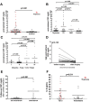
Results: Pretreatment CRP serum concentrations were significantly elevated in patients with TETs, particularly TCs and metastatic TETs. After complete tumor resection CRP serum concentrations were decreased (p = 0.135) but increased significantly in case of tumor recurrence (p = 0.001). High pretreatment CRP was associated with significantly worse 5- and 10-year freedom-from recurrence (FFR) (p = 0.010) and was a negative prognostic factor for FFR (HR 3.30; p = 0.015). IL-6 (not IL-1β) serum concentrations were significantly elevated in patients with TETs but we did not detect CRP tissue expression in TETs.
Materials and methods: Pretreatment CRP serum concentrations were retrospectively analyzed from 128 surgical patients (1990-2015). In a subset of 68 patients longitudinal analysis of CRP was performed. Additionally, immunohistochemical tumor CRP expression and serum concentrations of interleukin (IL)-6 and IL-1β were measured.
Conclusions: Hence, diagnostic measurement of serum CRP might be useful to indicate highly aggressive TETs and to make doctors consider tumor recurrences during oncological follow-up.
Keywords: CRP; prognosis; thymic carcinoma; thymic epithelial tumors; thymoma.
Conflict of interest statement
The authors declare no conflicts of interest
Figures


References
-
- Detterbeck FC, Nicholson AG, Kondo K, Van Schil P, Moran C. The Masaoka-Koga stage classification for thymic malignancies: clarification and definition of terms. J Thorac Oncol. 2011;6:1710–1716. - PubMed
-
- Kondo K, Yoshizawa K, Tsuyuguchi M, Kimura S, Sumitomo M, Morita J, Miyoshi T, Sakiyama S, Mukai K, Monden Y. WHO histologic classification is a prognostic indicator in thymoma. Ann Thorac Surg. 2004;77:1183–1188. - PubMed
-
- Ströbel P, Bauer A, Puppe B, Kraushaar T, Krein A, Toyka K, Gold R, Semik M, Kiefer R, Nix W, Schalke B, Müller-Hermelink HK, Marx A. Tumor recurrence and survival in patients treated for thymomas and thymic squamous cell carcinomas: a retrospective analysis. J Clin Oncol. 2004;22:1501–1509. - PubMed
-
- Moser B, Scharitzer M, Hacker S, Ankersmit J, Matilla JR, Lang G, Aigner C, Taghavi S, Klepetko W. Thymomas and thymic carcinomas: prognostic factors and multimodal management. Thorac Cardiovasc Surg. 2014;62:153–160. - PubMed
MeSH terms
Substances
Supplementary concepts
LinkOut - more resources
Full Text Sources
Other Literature Sources
Medical
Research Materials
Miscellaneous

