. 2017 Aug 1;313(2):R78-R90.
doi: 10.1152/ajpregu.00351.2016. Epub 2017 May 17.

Flexible ammonia handling strategies using both cutaneous and branchial epithelia in the highly ammonia-tolerant Pacific hagfish

Affiliations

Flexible ammonia handling strategies using both cutaneous and branchial epithelia in the highly ammonia-tolerant Pacific hagfish

Alexander M Clifford et al. Am J Physiol Regul Integr Comp Physiol. .

Abstract

Hagfish consume carrion, potentially exposing them to hypoxia, hypercapnia, and high environmental ammonia (HEA). We investigated branchial and cutaneous ammonia handling strategies by which Pacific hagfish (Eptatretus stoutii) tolerate and recover from high ammonia loading. Hagfish were exposed to HEA (20 mmol/l) for 48 h to elevate plasma total ammonia (TAmm) levels before placement into divided chambers for a 4-h recovery period in ammonia-free seawater where ammonia excretion (JAmm) was measured independently in the anterior and posterior compartments. Localized HEA exposures were also conducted by subjecting hagfish to HEA in either the anterior or posterior compartments. During recovery, HEA-exposed animals increased JAmm in both compartments, with the posterior compartment comprising ~20% of the total JAmm compared with ~11% in non-HEA-exposed fish. Plasma TAmm increased substantially when whole hagfish and the posterior regions were exposed to HEA. Alternatively, plasma TAmm did not elevate after anterior localized HEA exposure. JAmm was concentration dependent (0.05-5 mmol/l) across excised skin patches at up to eightfold greater rates than in skin sections that were excised from HEA-exposed hagfish. Skin excised from more posterior regions displayed greater JAmm than those from more anterior regions. Immunohistochemistry with hagfish-specific anti-rhesus glycoprotein type c (α-hRhcg; ammonia transporter) antibody was characterized by staining on the basal aspect of hagfish epidermis while Western blotting demonstrated greater expression of Rhcg in more posterior skin sections. We conclude that cutaneous Rhcg proteins are involved in cutaneous ammonia excretion by Pacific hagfish and that this mechanism could be particularly important during feeding.

Keywords: agnatha; cyclostome; nitrogen; rhesus glycoprotein; skin.


Figures

Fig. 1.
Fig. 1.
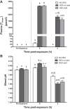
Changes in plasma total ammonia concentration ([TAmm]) (A) and blood pH (B) of Pacific hagfish exposed to high environmental ammonia (HEA) (closed bars; 20 mmol/l) for 48 h and after a 4-h recovery period in ammonia-free, full-strength seawater within divided chambers. Control fish (No HEA; open bars) were housed in ammonia-free seawater for the exposure period. Blood was drawn immediately before HEA exposure (Preexposure), after HEA exposure (0 h), and after a 4-h recovery period (4 h). After blood sampling was completed at 0 h, a subset of HEA-exposed hagfish were fitted with a cloacal seal (HEA seal; light gray bars), whereas the other group was left untreated (HEA no seal; dark gray bars) before being positioned into divided flux chambers. Data are presented as means + SE (n = 8) unless otherwise specified. Bars with same letter are not statistically different (P < 0.05) as determined by two-way ANOVA followed by Holm-Sidak’s multiple comparisons post hoc test with all possible comparisons made.
Fig. 2.
Fig. 2.
Partitioning of net outward ammonia excretion (JAmm) in Pacific hagfish exposed to HEA (20 mmol/l) for 48 h during recovery in ammonia-free, full-strength seawater within divided chambers. Control fish (No HEA; open bars) were housed in ammonia-free seawater for the duration of the exposure period. JAmm was measured in the anterior (A) and posterior (B) chambers during the 4-h recovery period. Data are presented as means + SE (n). Bars with same letter are not statistically different (P < 0.05) as determined by one-way ANOVA followed by Holm-Sidak’s multiple comparisons post hoc test. Details of cloacal seal application are described in Fig. 1.
Fig. 3.
Fig. 3.
Changes in plasma [TAmm] (A) and blood pH (B) during localized exposure of hagfish to HEA (20 mM) via either ammonia-free seawater (no HEA; open bars) or 20 mmol/l TAmm in either the anterior compartment (HEA in front) or the posterior compartment (HEA in back) divided chamber. Blood samples were drawn immediately after the 48-h exposure. Data are presented as means + SE (n). Bars with same letter are not statistically different (P < 0.05) as determined by a Kruskal-Wallis nonparametric test followed by Dunn’s multiple comparison test in A and by one-way ANOVA followed by Holm-Sidak’s multiple comparisons post hoc test in B.
Fig. 4.
Fig. 4.
Ammonia excretion during localized HEA exposure. Net outward anterior (A) and posterior (B) JAmm from hagfish during acute localized HEA exposure was determined in the compartment opposite to the HEA-containing compartment. Data are presented as means + SE (n). Bars with same letter are not statistically different (P < 0.05) as determined by Student’s one-tailed t-test.
Fig. 5.
Fig. 5.
Total ammonia flux across excised skin as determined by appearance of ammonia in mucosal medium following introduction to serosal HEA (0.05–10.0 mmol/l in hagfish saline). Concentration-dependent flux (A) was determined in excised skin from hagfish (n = 6) either preexposed to HEA (20 mmol/l) for 48 h (closed bars) or nonexposed controls (open bars). Skin JAmm was measured in skin excised from serial sections along the length of non-HEA-exposed animals. The serosal side of skin sections was then exposed to 5 mmol/l TAmm in hagfish saline, and the appearance of ammonia was measured on the mucosal side of the chamber. Data are presented as means + SE in A and means ± SE in B. Statistical differences in A (*P < 0.05) as determined by multiple Student’s t-tests with a Holm-Sidak multiple comparisons correction. Solid line in B represents line of best fit as determined by linear regression analysis {equation: skin JAmm = 0.1307 [(nmol·cm−2·h−1)/(% distance from snout)] + 18.82 nmol·cm−2·h−1}; broken lines represent 95% confidence limits for line of best fit, and the shaded area represents the placement of collar assembly on hagfish in separating chamber protocols.
Fig. 6.
Fig. 6.
Phylogenetic analysis of 2 cloned hagfish Rh glycoprotein sequences showed phylogenetic relationships of EsRhcg and EsRh-like peptide sequences. Analysis was completed using RAx-ML with methods previously described on the Cyberinfrastructure for Phylogenetic Research (CIPRES) Science Gateway servers. Branch support was estimated by bootstrap with 300 replications with auto-cutoff threshold set to 1,000. Cloned sequences are denoted with an arrow. Numbers in square brackets are the GenBank accession numbers of the sequences.
Fig. 7.
Fig. 7.
Representative micrographs depicting Pacific hagfish cutaneous cellular organization and EsRhcg localization. Hematoxylin and eosin staining (A) highlighting the large mucous cells (MC) and clearly defined basement membrane separating the epidermis (Ep) from the capillary-rich (Cp; denoted by arrows) dermis (De). Immunohistochemistry representative confocal images showing localization of the EsRhcg using Myxinid-derived antibodies (red) to the basal aspect of the epidermis (B). Immunoreactivity was observed along the length of the basement membrane of the epidermis, extending up to surround the large MC. Preabsorbed antibody control micrograph (B, inset) demonstrating no evidence of EsRhcg staining. Scale bars = 100 µm in A and 20 µm in B.
Fig. 8.
Fig. 8.
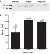
Cutaneous distribution of EsRhcg abundance in hagfish. Hagfish skin was excised in sequential sections (anterior, middle, posterior) from along the length of the animal. Skin sections were distributed by percentage distance from the snout (anterior: 27.58 ± 0.9%; middle: 57.21 ± 1.40%; posterior: 84.19 ± 1.08%). A: representative blot from anterior, middle, and posterior skin sections. B: relative abundance of cutaneous EsRhcg expression from sequential skin sections. Data are presented as means + SE (n). Bars with same letter are not statistically different (P < 0.05) as determined by one-way ANOVA followed by Holm-Sidak’s multiple comparisons post hoc test.

Comment in

  • Heads you gain, tails you lose.
    Gilmour KM. Gilmour KM. Am J Physiol Regul Integr Comp Physiol. 2017 Aug 1;313(2):R65-R66. doi: 10.1152/ajpregu.00208.2017. Epub 2017 Jun 7. Am J Physiol Regul Integr Comp Physiol. 2017. PMID: 28592460 Free PMC article. No abstract available.

References

    1. Alt JM, Stolte H, Eisenbach GM, Walvig F. Renal electrolyte and fluid excretion in the Atlantic hagfish Myxine glutinosa. J Exp Biol 91: 323–330, 1981.
    1. Bardack D. First fossil hagfish (myxinoidea): a record from the pennsylvanian of illinois. Science 254: 701–703, 1991. doi: 10.1126/science.254.5032.701. - DOI - PubMed
    1. Boutilier RG, Heming TA, Iwama GK. Appendix: physicochemical parameters for use in fish respiratory physiology In: Gills–Anatomy, Gas Transfer, and Acid-Base Regulation, edited by Hoar WS, Randall D. Orlando, FL: Elsevier, 1984, p. 403–430. doi: 10.1016/S1546-5098(08)60323-4. - DOI
    1. Braun MH, Perry SF. Ammonia and urea excretion in the Pacific hagfish Eptatretus stoutii: evidence for the involvement of Rh and UT proteins. Comp Biochem Physiol A Mol Integr Physiol 157: 405–415, 2010. doi: 10.1016/j.cbpa.2010.08.020. - DOI - PubMed
    1. Cameron JN, Heisler N. Studies of ammonia in the Rainbow trout: Physico-chemical parameters, acid-base behaviour and respiratory clearance. J Exp Biol 105: 107–125, 1983.

LinkOut - more resources