. 2017 Aug;66(8):2163-2174.
doi: 10.2337/db16-1285. Epub 2017 May 17.

Radiomanganese PET Detects Changes in Functional β-Cell Mass in Mouse Models of Diabetes

Affiliations

Radiomanganese PET Detects Changes in Functional β-Cell Mass in Mouse Models of Diabetes

Reinier Hernandez et al. Diabetes. 2017 Aug.

Abstract

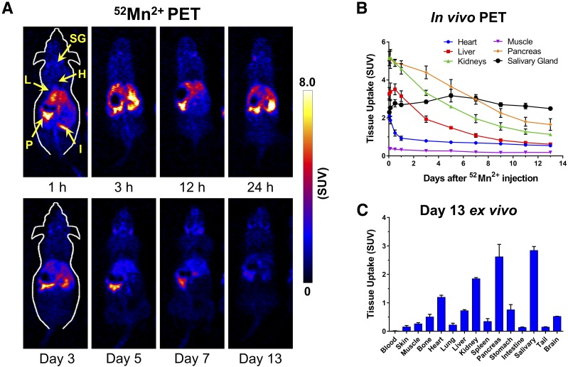
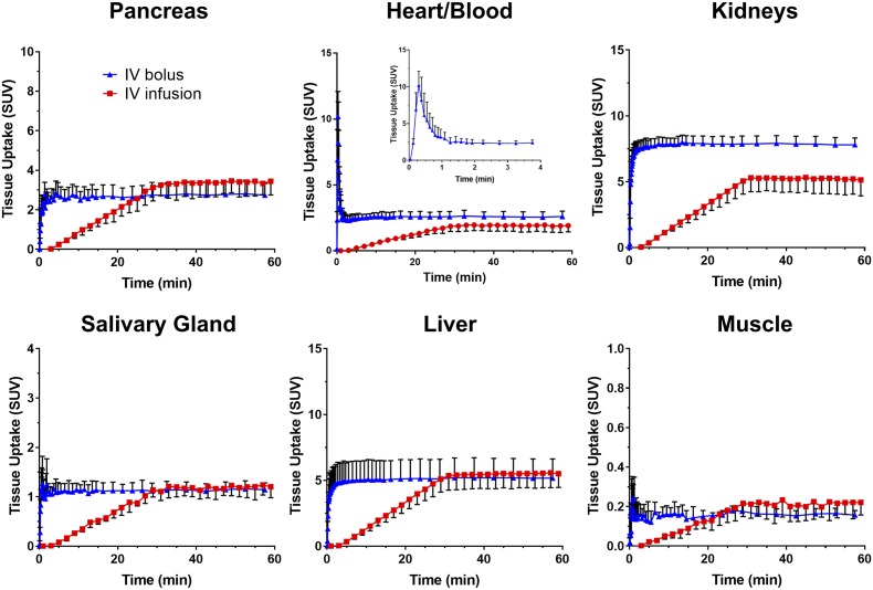
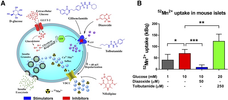
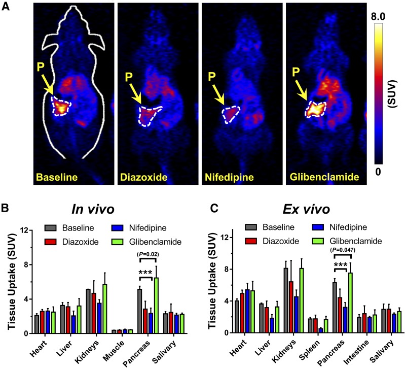
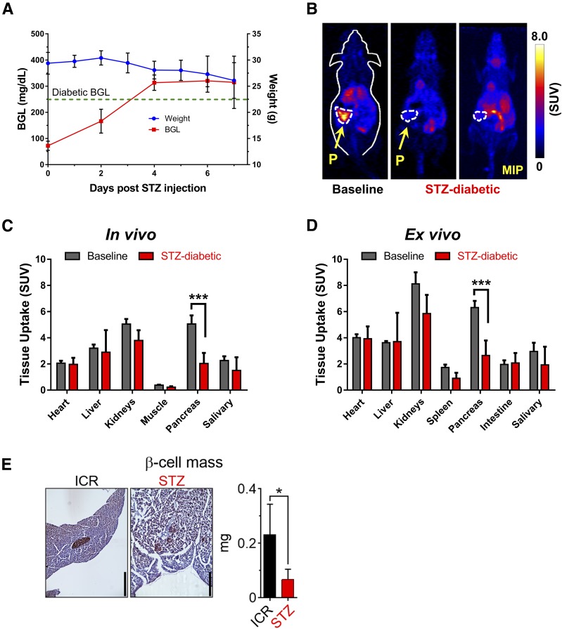
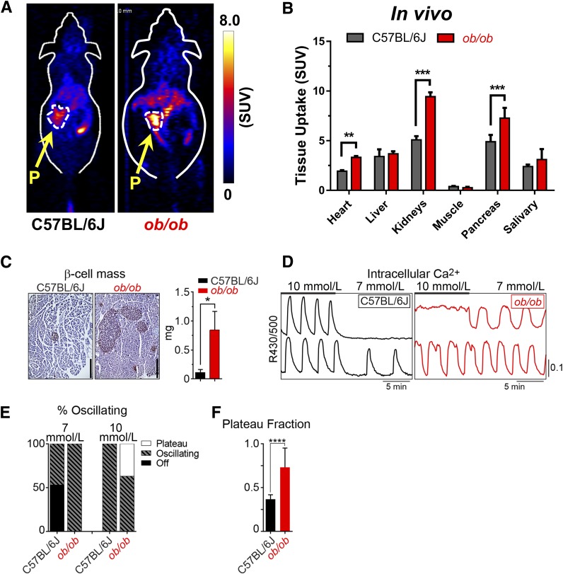
The noninvasive measurement of functional β-cell mass would be clinically valuable for monitoring the progression of type 1 and type 2 diabetes as well as the viability of transplanted insulin-producing cells. Although previous work using MRI has shown promise for functional β-cell mass determination through voltage-dependent Ca2+ channel (VDCC)-mediated internalization of Mn2+, the clinical utility of this technique is limited by the cytotoxic levels of the Mn2+ contrast agent. Here, we show that positron emission tomography (PET) is advantageous for determining functional β-cell mass using 52Mn2+ (t1/2: 5.6 days). We investigated the whole-body distribution of 52Mn2+ in healthy adult mice by dynamic and static PET imaging. Pancreatic VDCC uptake of 52Mn2+ was successfully manipulated pharmacologically in vitro and in vivo using glucose, nifedipine (VDCC blocker), the sulfonylureas tolbutamide and glibenclamide (KATP channel blockers), and diazoxide (KATP channel opener). In a mouse model of streptozotocin-induced type 1 diabetes, 52Mn2+ uptake in the pancreas was distinguished from healthy controls in parallel with classic histological quantification of β-cell mass from pancreatic sections. 52Mn2+-PET also reported the expected increase in functional β-cell mass in the ob/ob model of pretype 2 diabetes, a result corroborated by histological β-cell mass measurements and live-cell imaging of β-cell Ca2+ oscillations. These results indicate that 52Mn2+-PET is a sensitive new tool for the noninvasive assessment of functional β-cell mass.


Figures

Figure 1
Figure 1
Tissue distribution and pharmacokinetics of 52Mn2+. A: Serial PET images of ICR mice injected i.v. with 52Mn2+ (no anesthesia except during the PET scans). Coronal PET image slices were selected to best show pancreatic uptake. Arrows point to P, pancreas; H, heart; L, liver; I, intestines; and SG, salivary gland. B: ROI-based quantification of 52Mn2+ uptake in the heart, liver, kidneys, muscle, pancreas, and submandibular salivary gland. C: Ex vivo 52Mn2+ biodistribution of euthanized mice after the last PET scans, determined by gamma counting (n = 4).
Figure 2
Figure 2
Rapid kinetics of tissue 52Mn2+ uptake revealed by single i.v. bolus injection or continuous i.v. infusion. Dynamic PET TACs derived from hand-drawn ROIs for the pancreas, heart/blood, liver, kidneys, salivary gland, and muscle. The blue curves indicate TACs in mice injected with a rapid i.v. bolus of 52Mn2+, and the red curves indicate an i.v. infusion of 52Mn2+ over the first 30 min of the scans. The inset shows the blood/heart distribution of 52Mn2+ within the first 4 min after i.v. bolus injection.
Figure 3
Figure 3
Pharmacological manipulation of VDCC in isolated islets. A: Cartoon of the β-cell–triggering pathway. Molecular structures in blue indicate compounds that activate Ca2+ influx through VDCC, whereas compounds in red are inhibitory. B: Uptake of 52Mn2+ by isolated ob/ob mouse islets. Groups of 50 islets from three preparations were incubated with 52Mn2+ (370 kBq) in the presence of glucose and VDCC modulators as indicated. Data are presented as mean ± SD. *P < 0.05; **P < 0.01; ***P < 0.001.
Figure 4
Figure 4
In vivo assessment of functional β-cell mass by 52Mn2+-PET. A: Coronal PET images at 1 h postinjection showing the pancreas of ICR mice given i.p. injections of diazoxide (20 mg/kg), nifedipine (20 mg/kg), or glibenclamide (5 mg/kg) before the administration of a 52Mn2+ rapid bolus. The pancreas (P) is demarcated by white dashed contours. B: Manual ROI-based quantification of 52Mn2+ uptake in various tissues from static PET images acquired at 1 h postinjection. C: Ex vivo biodistribution analysis after PET imaging at 1 h postinjection. Significantly reduced pancreatic uptake of 52Mn2+ is observed in mice that received nifedipine and diazoxide before radiotracer administration. Mice that received glibenclamide (5 mg/kg) before radiotracer administration had significantly higher pancreatic uptake of 52Mn2+ than the control mice, based on both PET imaging (P = 0.02) and biodistribution (P = 0.047) studies. Data are presented as mean ± SD (n = 3–4 mice per group). ***P < 0.001.
Figure 5
Figure 5
52Mn2+-PET imaging in STZ-induced type 1 diabetes. A: After the administration of an acute dose of STZ (180 mg/kg), ICR mice started to show symptoms of diabetes: reduced body weight and high blood glucose level (BGL; >250 mg/dL). B: At 1 h postinjection, coronal PET images of healthy (left panel) or diabetic (center/right panels) ICR mice show clearly reduced PET signal in the pancreas of the STZ-diabetic mice. The pancreas (P) is demarcated by white dashed contours. The significant decline in 52Mn2+ uptake in the pancreas of STZ-diabetic mice was confirmed quantitatively by ROI analysis of the PET images (C) and ex vivo biodistribution (D) (n = 3 mice/group). E: Quantification of β-cell mass for control and STZ-treated ICR mice. Data are shown as the mean ± SD (n = 4 mice/group). Scale bars = 400 μm. *P < 0.05, ***P < 0.001. MIP, maximum intensity projection.
Figure 6
Figure 6
52Mn2+-PET imaging in the ob/ob model of pretype 2 diabetes. A: Coronal PET images acquired at 1 h after 52Mn2+ administration in ob/ob mice and C57BL/6J controls. The pancreas (P) is demarcated by white dashed contours. B: Image-derived quantification expressed as SUV indicated a significant difference in 52Mn2+ pancreatic uptake between groups (mean ± SD; n = 3). C: Quantification of β-cell mass for wild-type and ob/ob mice (n = 3 mice/group). Scale bars = 400 μm. D: Recordings of islet Ca2+ in response to glucose (10 mmol/L and 7 mmol/L) from wild-type and ob/ob C57BL/6J mice. E: Contingency plot shows the range of islet behaviors at each glucose level. F: The oscillatory plateau fraction, reflecting the plasma membrane glucose sensitivity, was calculated as the fraction of time spent in the active state during each oscillation at 10 mmol/L glucose. C57BL/6J, n = 141 islets from 4 mice; ob/ob, n = 126 islets from 4 mice. Results reflect mean ± SD. *P < 0.05, **P < 0.01, ***P < 0.001, ****P < 0.0001.

References

    1. Forbes JM, Cooper ME. Mechanisms of diabetic complications. Physiol Rev 2013;93:137–188 - PubMed
    1. Ionescu-Tirgoviste C, Gagniuc PA, Gubceac E, et al. A 3D map of the islet routes throughout the healthy human pancreas. Sci Rep 2015;5:14634. - PMC - PubMed
    1. Kim A, Miller K, Jo J, Kilimnik G, Wojcik P, Hara M. Islet architecture: a comparative study. Islets 2009;1:129–136 - PMC - PubMed
    1. von Mach MA, Schlosser J, Weiland M, et al. Size of pancreatic islets of Langerhans: a key parameter for viability after cryopreservation. Acta Diabetol 2003;40:123–129 - PubMed
    1. Kilimnik G, Jo J, Periwal V, Zielinski MC, Hara M. Quantification of islet size and architecture. Islets 2012;4:167–172 - PMC - PubMed

Publication types

MeSH terms