. 2017 Jul;206(3):1521-1534.
doi: 10.1534/genetics.116.195297. Epub 2017 May 17.

Regulation of WNT Signaling at the Neuromuscular Junction by the Immunoglobulin Superfamily Protein RIG-3 in Caenorhabditis elegans

Affiliations

Regulation of WNT Signaling at the Neuromuscular Junction by the Immunoglobulin Superfamily Protein RIG-3 in Caenorhabditis elegans

Pratima Pandey et al. Genetics. 2017 Jul.

Abstract

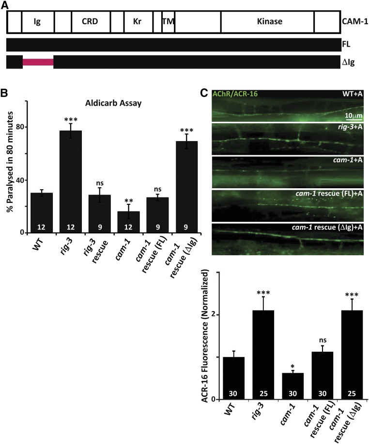
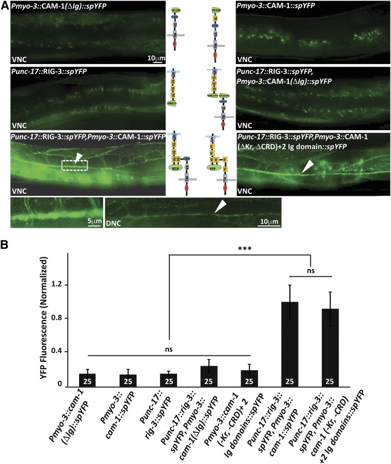
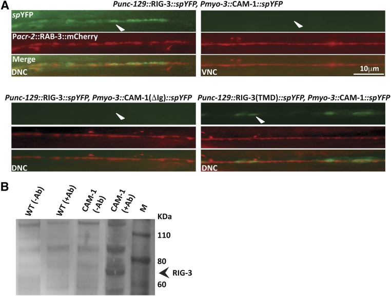
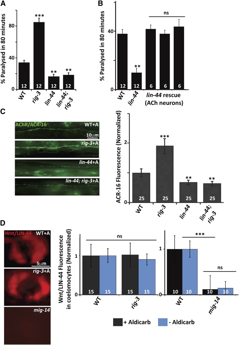
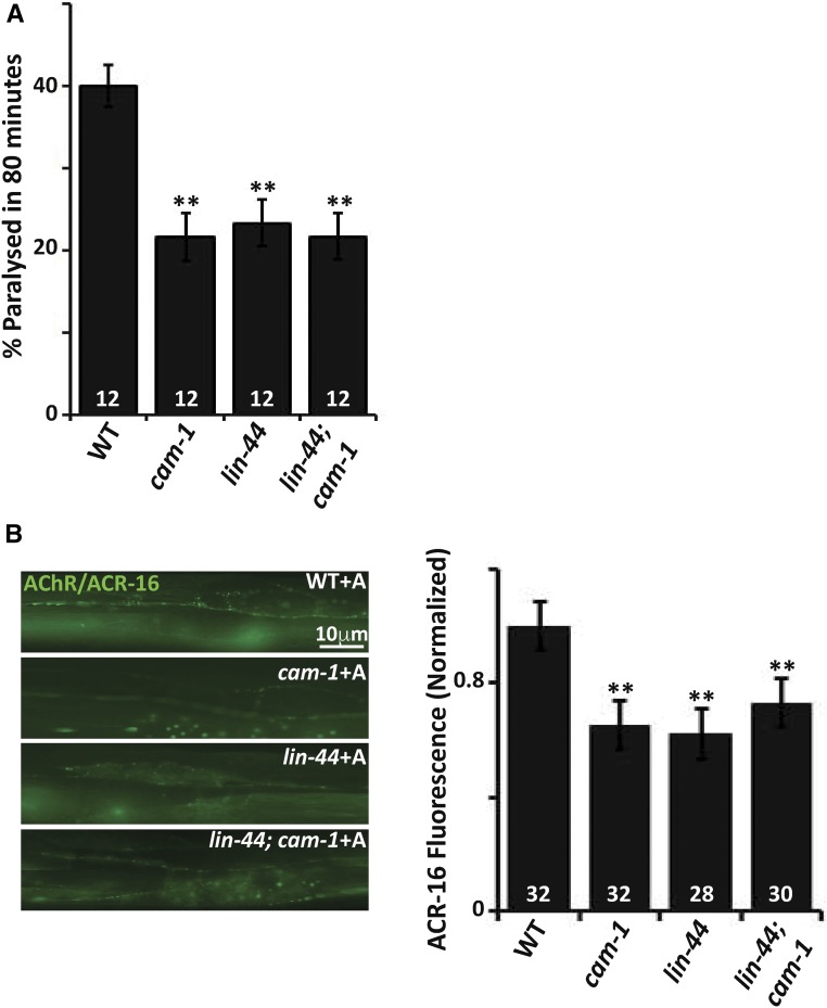
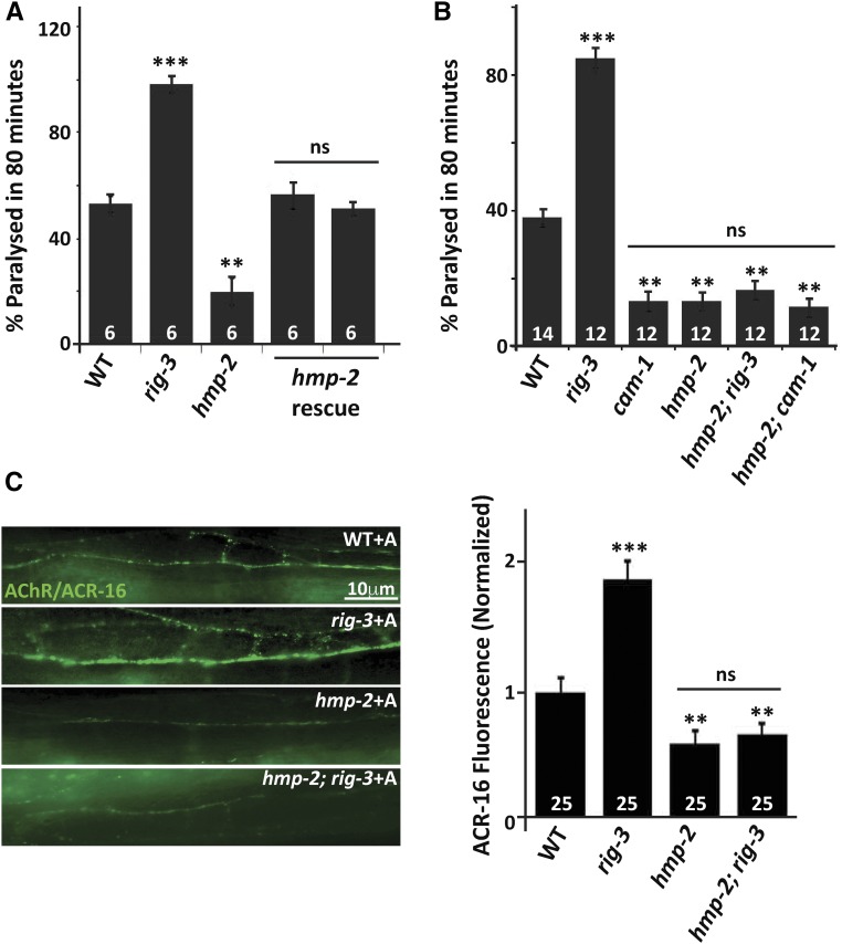
Perturbations in synaptic function could affect the normal behavior of an animal, making it important to understand the regulatory mechanisms of synaptic signaling. Previous work has shown that in Caenorhabditis elegans an immunoglobulin superfamily protein, RIG-3, functions in presynaptic neurons to maintain normal acetylcholine receptor levels at the neuromuscular junction (NMJ). In this study, we elucidate the molecular and functional mechanism of RIG-3. We demonstrate by genetic and BiFC (Bi-molecular Fluorescence Complementation) assays that presynaptic RIG-3 functions by directly interacting with the immunoglobulin domain of the nonconventional Wnt receptor, ROR receptor tyrosine kinase (RTK), CAM-1, which functions in postsynaptic body-wall muscles. This interaction in turn inhibits Wnt/LIN-44 signaling through the ROR/CAM-1 receptor, and allows for maintenance of normal acetylcholine receptor, AChR/ACR-16, levels at the neuromuscular synapse. Further, this work reveals that RIG-3 and ROR/CAM-1 function through the β-catenin/HMP-2 at the NMJ. Taken together, our results demonstrate that RIG-3 functions as an inhibitory molecule of the Wnt/LIN-44 signaling pathway through the RTK, CAM-1.

Keywords: C. elegans; RIG-3; Wnt; immunoglobulin domain; neuromuscular junction.


Figures

Figure 1
Figure 1
RIG-3 functions through the Ig domain of CAM-1. (A) Schematic of CAM-1: the panel indicates schematic representations of CAM-1 domains for Full Length (FL) CAM-1, and the CAM-1 coding sequence lacking the Immunoglobulin domain, i.e., CAM-1(ΔIg). CRD, Cysteine Rich Domain; Kr, Kringle domain; TM, Transmembrane domain. (B) Aldicarb assays of rig-3 and cam-1 mutants and their rescue lines: this panel indicates a bar- graph showing the percentage of C. elegans paralyzed on aldicarb at the end of 80 min. In all figures, mutant and rescue lines were tested against control WT animals for significant differences. * P < 0.05, ** P < 0.01, and *** P < 0.001. The numbers at the base of the bars for all figures showing aldicarb assays indicates number of times the assay was performed with 15–25 animals used for each trial. (C) Quantitation of AChR/ACR-16 in rig-3 and cam-1 mutants along with the CAM-1 rescue lines: the top portion of the panel shows images of AChR/ACR-16 tagged with GFP in control WT, rig-3, and cam-1 mutants, and CAM-1(FL) and CAM-1(ΔIg) rescue lines. The lower part of the panel shows quantitation of AChR/ACR-16 levels along the Ventral Nerve Cord (VNC). The numbers analyzed for each genotype are indicated. In all figures “+A” indicates “+Aldicarb,” where 1 mM aldicarb treatment was done for 80 min unless otherwise mentioned. In (B and C), the CAM-1 rescue experiments were done by expressing CAM-1 specifically in the body-wall muscle with the myo-3 promoter. In (B) and (C), RIG-3 rescue was done by expressing RIG-3 under it’s own promoter, which is expressed in cholinergic motor neurons. Error bars indicate SEM in all panels.
Figure 2
Figure 2
RIG-3 directly interacts with CAM-1. (A) Bimolecular fluorescence complementation (BiFC) analysis of CAM-1 and RIG-3 interaction at the NMJ: the figure shows representative fluorescent images (YFP) of the C. elegans Dorsal Nerve Cord (DNC) expressing the transgenic protein indicated. The top four panels display the controls showing basal YFP levels with Pmyo-3::CAM-1(ΔIg)::VC155, Pmyo-3::CAM-1(FL)::VC155, Punc-17::RIG-3::VN173, and Punc-17::RIG-3::VN173 coinjected with Pmyo-3::CAM-1(ΔIg)::VC155. The lower two panels that are adjacent to the illustrations of the constructs used in the BiFC experiments show the image where Punc-17::RIG-3::VN173 is coinjected with Pmyo-3::CAM-1(FL)::VC155 and Punc-17::RIG-3::VN173 is coinjected with Pmyo-3::CAM-1(ΔKr, ΔCRD)+ 2Ig::VC155 respectively. A clear increase over the basal levels of YFP is seen in these panels. The image to the bottom right of the figure is a small area along the VNC that has been zoomed into from the image on the left (dotted box). The image next to it shows expression of YFP along the DNC in the same animals that show YFP expression in the VNC, this was seen in all eight animals that were assayed to look at the DNC. VNC indicates ventral nerve cord, and DNC indicates dorsal nerve cord, respectively. CAM-1 was expressed in the body-wall muscle with the myo-3 promoter, while RIG-3 was expressed in cholinergic neurons using the unc-17 promoter. (B) Graph of the bimolecular fluorescence images: In order to quantify the YFP fluorescence levels, 25 animals for each genotype where imaged, and their fluorescence along the ventral nerve cord quantified and plotted in the graph. The fluorescence intensity was normalized against C. elegans coexpressing Punc-17::RIG-3::VN173 and Pmyo-3::CAM-1(FL)::VC155. Error bars indicate SEM.
Figure 3
Figure 3
Interaction between RIG-3 and CAM-1. (A) YFP reconstitution across the dorsal cord in the background of Pacr-2::mCherry::RAB-3 that shows expression along both the cords: RIG-3 expressed under the unc-129 promoter interacts with CAM-1 expressed in body-wall muscle (myo-3 promoter) along the dorsal cord, but not along the ventral cord (top rows; arrowheads). The lower left set of images show the control, where RIG-3 does not interact with CAM-1(ΔIg). RIG-3(TMD) also shows interaction with CAM-1 along the dorsal cord (lower right panels, arrowhead). (B) Coimmunoprecipitation experiment: the panel shows the coimmunoprecipitation (Co-IP) blot. Protein extracts isolated from WT and CAM-1::GFP animals were allowed to interact with GFP antibody and pulled down using ProteinA/G beads. As a control, the same protein extracts were allowed to bind to beads but without GFP antibody. The coimmunoprecipitated complex was probed with the anti-RIG-3 antibody. The RIG-3 containing complex was pulled down only in the lane containing the CAM-1::GFP animals, and not in any of the other three control lanes (arrowhead). Co-IP analysis indicates interaction between CAM-1 and RIG-3.
Figure 4
Figure 4
RIG-3 functions through the Wnt/LIN-44 at the NMJ. (A) Aldicarb assay for Wnt/lin-44; rig-3 mutants, and control strains: paralysis on aldicarb was assayed in WT control animals, rig-3, Wnt/lin-44, and Wnt/lin-44; rig-3 double mutants. (B) Rescue of the Wnt/lin-44 mutant phenotype: Graph indicates rescue of the aldicarb resistance phenotype seen in the Wnt/lin-44 mutants. The rescue constructs of all three rescuing lines expressed Wnt/LIN-44 in the cholinergic neurons using the unc-17 promoter. The three bars indicate three independent rescuing lines of Wnt/LIN-44. (C) The increased AChR/ACR-16 expression seen in rig-3 mutants is suppressed by Wnt/lin-44 mutation: the left panel displays images of WT, rig-3, Wnt/lin-44, and Wnt/lin-44; rig-3 double mutants showing expression of AChR/ACR-16 tagged with GFP along the VNC of C. elegans after treating the animals with aldicarb. The right side of the panel shows a graph of AChR/ACR-16 quantitation, which has been normalized against the average AChR/ACR-16 fluorescence values in WT animals. (D) RIG-3 does not affect Wnt/LIN-44 secretion: the left side of the panel shows the images of coelomocytes with Wnt/LIN-44 tagged to the mCherry marker in WT, rig-3 and mig-14 mutants. The coelomocytes show uptake of the Wnt/LIN-44 that has been secreted from cholinergic neurons where it was originally expressed. The right side of the panel shows the quantitation of the coelomocyte fluorescence normalized to WT. The black bars indicate normalized coelomocyte fluorescence in WT and rig-3 mutants before aldicarb treatment, while the blue bars indicate the normalized fluorescent values after aldicarb treatment. The number at the base of the graph indicates the number of animals that were tested. The second bar shows a similar quantitation where the coelomocyte fluorescence is compared between WT and mig-14 animals. Error bars indicate SEM in all panels.
Figure 5
Figure 5
Wnt/LIN-44 could function through CAM-1 at the NMJ. (A) Wnt/LIN-44 and CAM-1 genetically interact with each other: the graph indicates paralysis on aldicarb for Wnt/lin-44; cam-1 double mutants and control animals. The assay was performed on 1 mM aldicarb containing plates. The percentage of animals that paralyzed at 80 min were plotted, and statistical analysis was performed to ascertain significant difference of mutant values from the WT C. elegans. (B) Wnt/LIN-44 could function through CAM-1 to maintain AChR/ACR-16 levels at the NMJ: the left side of the panel shows images of AChR/ACR-16 tagged with GFP along the VNC. The right side of the panel shows the quantitation of the AChR/ACR-16 fluorescence levels that are normalized to control WT fluorescent values. Error bars indicate SEM in all panels.
Figure 6
Figure 6
RIG-3 functions through the β-catenin/HMP-2 at the C. elegans NMJ. (A) The resistance to aldicarb phenotype seen in β−catenin/hmp-2 mutants is rescued by expressing β−Catenin/HMP-2 in the body-wall muscle: This panel shows a graph of aldicarb induced paralysis shown for WT, rig-3 controls, β−catenin/hmp-2 mutants, and rescue of β−catenin/hmp-2 by expressing HMP-2 in the body-wall muscles of C. elegans using the myo-3 promoter. The HMP-2 rescue was done using two independent array lines, and data from both these lines are represented in this panel as separate bars. (B) Both RIG-3 and CAM-1 function through the β-catenin/HMP-2 at the NMJ: this panel shows a graph of aldicarb induced paralysis shown for β−catenin/hmp-2; rig-3 and β−catenin/hmp-2; cam-1 double mutants with controls. (C) The β−catenin/hmp-2 mutant shows decreased AChR/ACR-16 levels: this panel shows decreased AChR/ACR-16 in β−catenin/hmp-2 mutants along the VNC after treatment of the animals with aldicarb. WT and rig-3 mutants treated with aldicarb are shown as controls. Error bars indicate SEM in all panels.

Similar articles

Cited by

References

    1. Ackley B. D., 2014. Wnt-signaling and planar cell polarity genes regulate axon guidance along the anteroposterior axis in C. elegans. Dev. Neurobiol. 74: 781–796. - PMC - PubMed
    1. Babu K., Hu Z., Chien S. C., Garriga G., Kaplan J. M., 2011. The immunoglobulin super family protein RIG-3 prevents synaptic potentiation and regulates Wnt signaling. Neuron 71: 103–116. - PMC - PubMed
    1. Bei Y., Hogan J., Berkowitz L. A., Soto M., Rocheleau C. E., et al. , 2002. SRC-1 and Wnt signaling act together to specify endoderm and to control cleavage orientation in early C. elegans embryos. Dev. Cell 3: 113–125. - PubMed
    1. Bielen H., Houart C., 2014. The Wnt cries many: Wnt regulation of neurogenesis through tissue patterning, proliferation, and asymmetric cell division. Dev. Neurobiol. 74: 772–780. - PubMed
    1. Bilimoria P. M., Bonni A., 2013. Molecular control of axon branching. Neuroscientist 19: 16–24. - PMC - PubMed

Publication types

MeSH terms