. 2017 Jul 12;91(15):e00528-17.
doi: 10.1128/JVI.00528-17. Print 2017 Aug 1.

Influenza A Virus NS1 Protein Promotes Efficient Nuclear Export of Unspliced Viral M1 mRNA

Affiliations

Influenza A Virus NS1 Protein Promotes Efficient Nuclear Export of Unspliced Viral M1 mRNA

Carina F Pereira et al. J Virol. .

Abstract

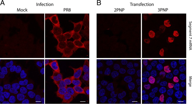
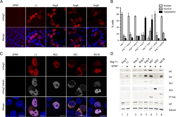
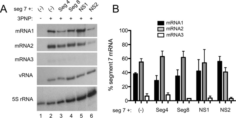
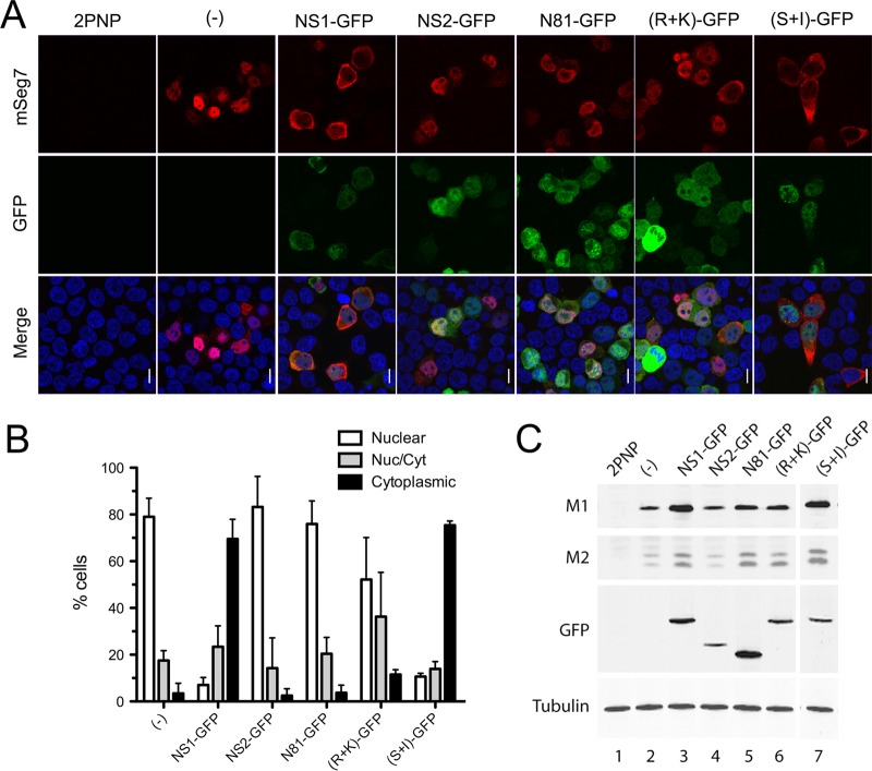
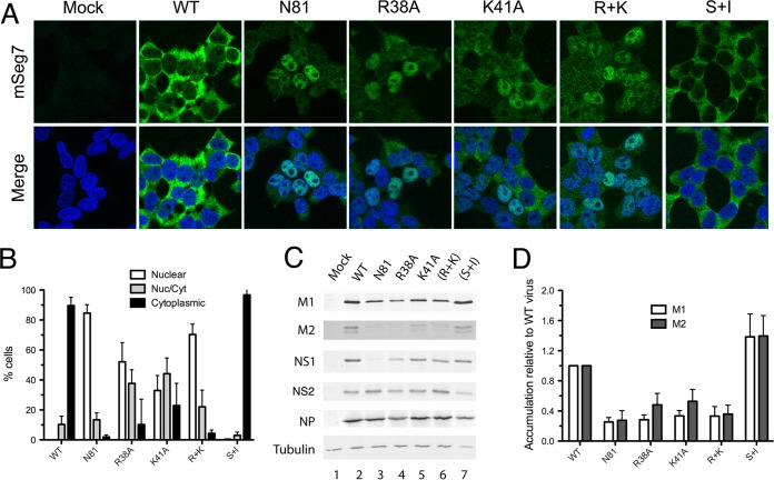
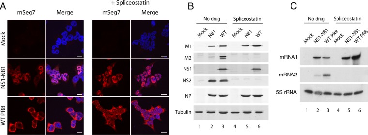
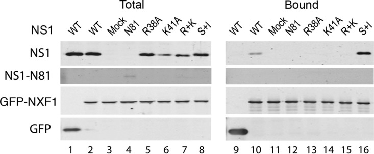
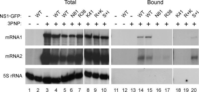
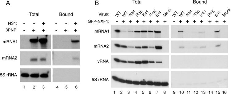
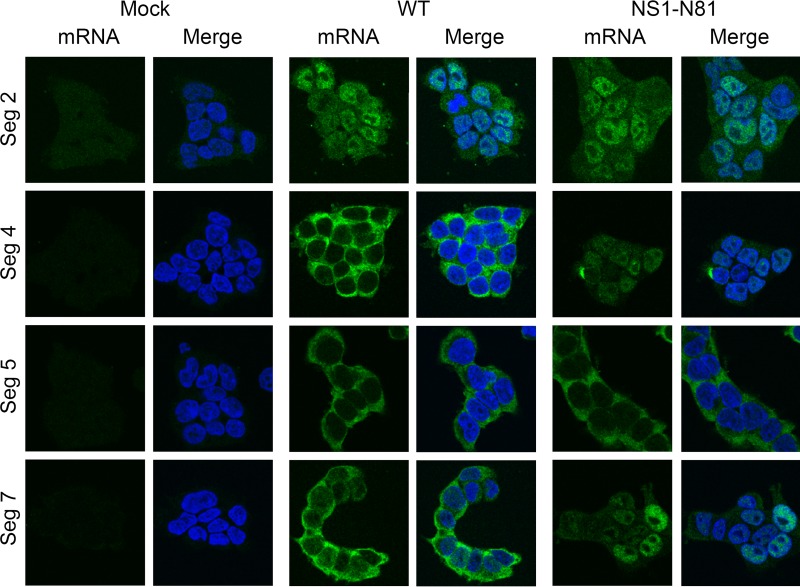
Influenza A virus mRNAs are transcribed by the viral RNA-dependent RNA polymerase in the cell nucleus before being exported to the cytoplasm for translation. Segment 7 produces two major transcripts: an unspliced mRNA that encodes the M1 matrix protein and a spliced transcript that encodes the M2 ion channel. Export of both mRNAs is dependent on the cellular NXF1/TAP pathway, but it is unclear how they are recruited to the export machinery or how the intron-containing but unspliced M1 mRNA bypasses the normal quality-control checkpoints. Using fluorescent in situ hybridization to monitor segment 7 mRNA localization, we found that cytoplasmic accumulation of unspliced M1 mRNA was inefficient in the absence of NS1, both in the context of segment 7 RNPs reconstituted by plasmid transfection and in mutant virus-infected cells. This effect was independent of any major effect on steady-state levels of segment 7 mRNA or splicing but corresponded to a ∼5-fold reduction in the accumulation of M1. A similar defect in intronless hemagglutinin (HA) mRNA nuclear export was seen with an NS1 mutant virus. Efficient export of M1 mRNA required both an intact NS1 RNA-binding domain and effector domain. Furthermore, while wild-type NS1 interacted with cellular NXF1 and also increased the interaction of segment 7 mRNA with NXF1, mutant NS1 polypeptides unable to promote mRNA export did neither. Thus, we propose that NS1 facilitates late viral gene expression by acting as an adaptor between viral mRNAs and the cellular nuclear export machinery to promote their nuclear export.IMPORTANCE Influenza A virus is a major pathogen of a wide variety of mammalian and avian species that threatens public health and food security. A fuller understanding of the virus life cycle is important to aid control strategies. The virus has a small genome that encodes relatively few proteins that are often multifunctional. Here, we characterize a new function for the NS1 protein, showing that, as well as previously identified roles in antagonizing the innate immune defenses of the cell and directly upregulating translation of viral mRNAs, it also promotes the nuclear export of the viral late gene mRNAs by acting as an adaptor between the viral mRNAs and the cellular mRNA nuclear export machinery.

Keywords: NS1; NXF1; influenza A virus mRNA export; nuclear import/export.


Figures

FIG 1
FIG 1
Localization of segment 7 mRNA in infected and transfected cells. 293T cells were infected or mock infected with Cambridge PR8 at an MOI of 5 and fixed at 6 h p.i. (A) or transfected with plasmids to reconstitute RNPs (3PNP) containing segment 7 vRNA or with a negative-control set lacking PB2 (2PNP) and fixed 24 h later before. Cells were then stained for positive-sense segment 7 RNA by FISH (red) or for DNA (4′,6′-diamidino-2-phenylindole; blue) and imaged by confocal microscopy (B). Single optical slices are shown. Scale bar, 10 μm.
FIG 2
FIG 2
NS1 promotes cytoplasmic accumulation of M1 mRNA. 293T cells were transfected with plasmids to reconstitute RNPs (3PNP) containing segment 7 vRNA or with a negative-control set lacking PB2 (2PNP or −) as well as with other segments or plasmids expressing NS1 or NS2 only or NS1 from influenza B virus (NS1B) and fixed 24 h later. Cells were then stained for positive-sense segment 7 RNA (red) (A and C) or the intronic sequence of M1 (gray) by FISH or for DNA (4′,6′-diamidino-2-phenylindole; blue) and imaged by confocal microscopy (C). Single optical slices are shown. Scale bar, 10 μm. (B) Individual cells were scored as to whether segment 7 mRNA staining was predominantly nuclear, cytoplasmic, or mixed. The means ± standard errors of the means from two to five independent experiments are plotted. (D) Cell lysates were analyzed by Western blotting for the indicated antigens. mSeg7, segment 7 mRNA.
FIG 3
FIG 3
Accumulation of segment 7 mRNA species in RNP reconstitution assays. (A) 293T cells were transfected with plasmids to recreate segment 7 RNPs or, as a negative control, with a 2PNP combination lacking PB2 either alone (−) or along with other viral components as labeled. Twenty-four hours later, total cellular RNA was extracted and analyzed by radioactive reverse transcriptase primer extension followed by urea-PAGE and autoradiography with primers specific for segment 7 mRNAs or cellular 5S rRNA. (B) Replicate experiments were quantified by densitometry, and the percentage of total segment 7 mRNA for each of the three species was determined. Data are the means ± standard deviations (n = 3).
FIG 4
FIG 4
Ability of GFP-tagged NS polypeptides to support segment 7 mRNA export. (A) 293T cells were transfected with plasmids to reconstitute RNPs containing segment 7 vRNA or with a negative-control set lacking PB2 (2PNP) as well as with plasmids expressing the indicated GFP-tagged proteins and fixed 24 h later before being stained for GFP (green), positive-sense segment 7 RNA (red, and DNA (4′,6′-diamidino-2-phenylindole; blue) and imaged by confocal microscopy. Single optical slices are shown. Scale bar, 10 μm. (B) Individual cells were scored as to whether segment 7 mRNA staining was predominantly nuclear, cytoplasmic, or mixed. Values are the means ± standard deviations from two to five independent experiments. (C) Parallel samples were processed by Western blotting for the indicated polypeptides.
FIG 5
FIG 5
Effect of NS1 mutations on segment 7 mRNA localization in infected cells. (A) 293T cells were infected with the indicated viruses at an MOI of 5, and at 6 h p.i. they were stained for segment 7 mRNA by FISH (green) and for DNA (4′,6′-diamidino-2-phenylindole; blue) before confocal imaging. Single optical slices are shown. Scale bar, 10 μm. (B) Individual cells were scored as to whether segment 7 mRNA staining was predominantly nuclear, cytoplasmic, or mixed. Values are the means ± standard errors of the means from three to six independent experiments. (C) Cell lysates were analyzed by Western blotting for the indicated antigens. (D) M1 and M2 accumulation from replicate experiments was quantified and expressed as a ratio relative to NP expression. Values plotted are normalized to the ratio seen with WT virus and are the means ± standard errors of the means of three independent experiments.
FIG 6
FIG 6
Effect of spliceostatin A on segment 7 mRNA localization. (A) 293T cells were infected with reverse genetics PR8 virus at an MOI of 5 and at 6 h p.i. treated or mock treated with 100 μg/ml spliceostatin A. At 6 h p.i. samples were fixed and stained by FISH for segment 7 mRNA (red) or by 4′,6′-diamidino-2-phenylindole for DNA (blue) and imaged by confocal microscopy. Single optical slices are shown. Scale bar, 10 μm. (B) Cell lysates were examined by Western blotting for the indicated polypeptides. (C) Total cellular RNA was analyzed by radioactive primer extension, urea-PAGE, and autoradiography for the indicated RNA species.
FIG 7
FIG 7
Interaction between NXF1 and NS1. 293T cells were transfected with plasmids encoding GFP or GFP-NXF1 and 48 h later either mock infected or infected with the indicated NS1 mutant viruses. Cells were harvested at 6 h p.i., and cell lysates were examined by Western blotting for the indicated proteins before (Total) or after (Bound) fractionation over GFP-Trap agarose.
FIG 8
FIG 8
Interaction between segment 7 mRNAs and NS1. 293T cells were transfected with plasmids to recreate segment 7 RNPs (3PNP +) or, as a negative control, with a 2PNP combination (3PNP −) along with the indicated GFP-NS1 polypeptides or with GFP only (NS1-GFP −). Forty-eight hours later, total cellular RNA was extracted and analyzed by radioactive reverse transcriptase primer extension followed by urea-PAGE and autoradiography with primers specific for segment 7 mRNAs or cellular 5S rRNA.
FIG 9
FIG 9
NS1 promotes the interaction of segment 7 mRNA with NXF1. (A) 293T cells were transfected with plasmids to recreate segment 7 RNPs (3PNP +) or, as a negative control, with a 2PNP combination (3PNP −) along with GFP-NXF1 and with or without NS1 as labeled. Forty-eight hours later, total cellular RNA was extracted and analyzed by radioactive reverse transcriptase primer extension followed by urea-PAGE and autoradiography with primers specific for segment 7 mRNAs or cellular 5S rRNA. (B) Cells were transfected with GFP-NXF1 (+) or with GFP alone (−) and 48 h later infected or mock infected with the indicated viruses at an MOI of 10. Total RNA was extracted at 6 h p.i. and analyzed as described for panel A except that a primer specific for segment 7 vRNA was also included.
FIG 10
FIG 10
Role of NS1 in promoting export of other viral mRNAs. 293T cells were infected or mock infected with the indicated reverse genetics PR8 viruses at an MOI of 5, fixed at 6 h p.i., and stained for positive-sense RNAs from the indicated segments (green) and for DNA (4′,6′-diamidino-2-phenylindole; blue) before confocal imaging. Single optical slices are shown. Scale bar, 10 μm.

References

    1. Te Velthuis AJ, Fodor E. 2016. Influenza virus RNA polymerase: insights into the mechanisms of viral RNA synthesis. Nat Rev Microbiol 14:479–493. doi: 10.1038/nrmicro.2016.87. - DOI - PMC - PubMed
    1. Hutchinson EC, Fodor E. 2013. Transport of the influenza virus genome from nucleus to nucleus. Viruses 5:2424–2446. doi: 10.3390/v5102424. - DOI - PMC - PubMed
    1. Boulo S, Akarsu H, Ruigrok RW, Baudin F. 2007. Nuclear traffic of influenza virus proteins and ribonucleoprotein complexes. Virus Res 124:12–21. doi: 10.1016/j.virusres.2006.09.013. - DOI - PubMed
    1. Dubois J, Terrier O, Rosa-Calatrava M. 2014. Influenza viruses and mRNA splicing: doing more with less. mBio 5:e00070-14. doi: 10.1128/mBio.00070-14. - DOI - PMC - PubMed
    1. Inglis SC, Carroll AR, Lamb RA, Mahy BW. 1976. Polypeptides specified by the influenza virus genome. I. Evidence for eight distinct gene products specified by fowl plague virus. Virology 74:489–503. - PubMed

MeSH terms

LinkOut - more resources