. 2017 May 30;114(22):E4370-E4379.
doi: 10.1073/pnas.1705898114. Epub 2017 May 17.

Control of Hsp90 chaperone and its clients by N-terminal acetylation and the N-end rule pathway

Affiliations

Control of Hsp90 chaperone and its clients by N-terminal acetylation and the N-end rule pathway

Jang-Hyun Oh et al. Proc Natl Acad Sci U S A. .

Abstract

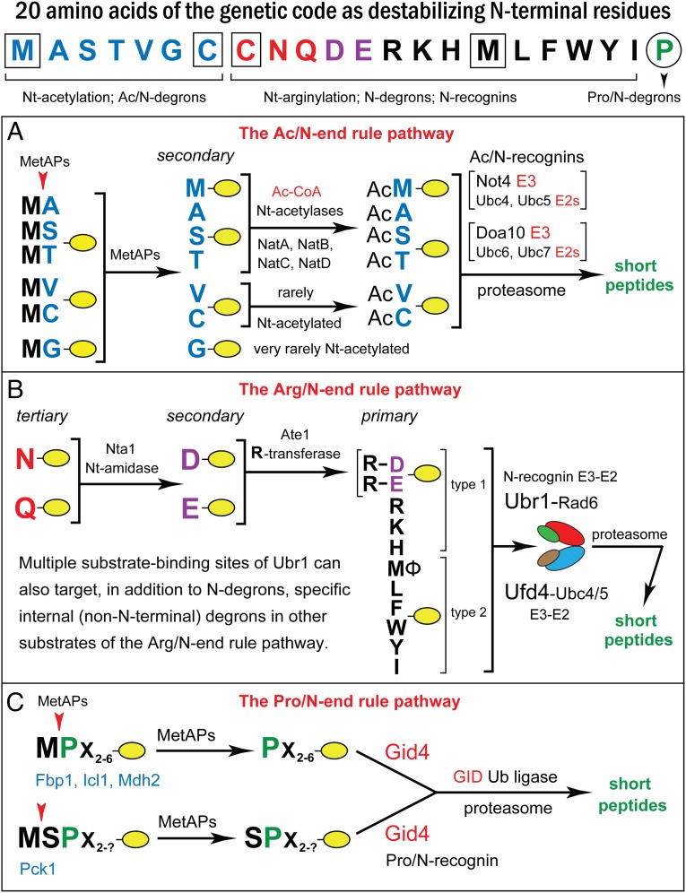
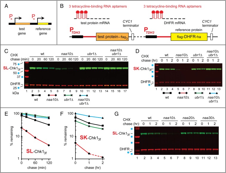
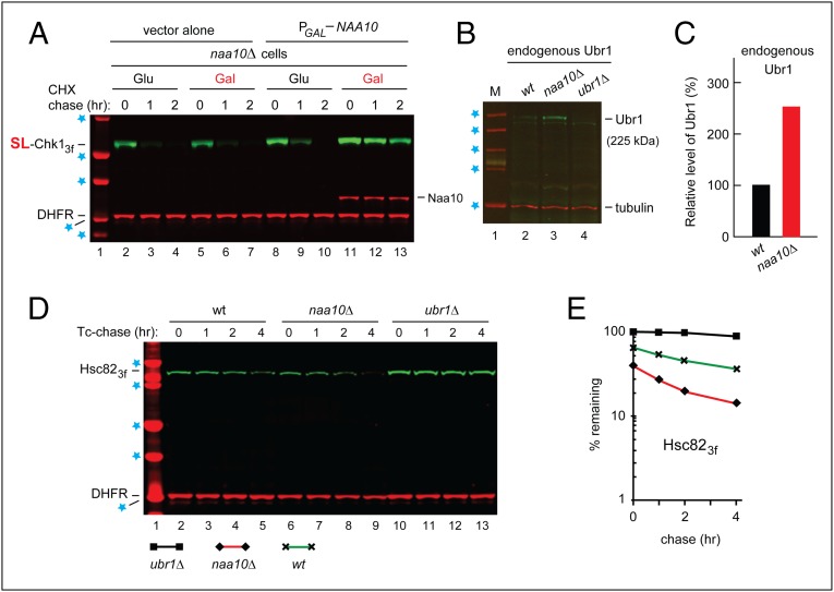
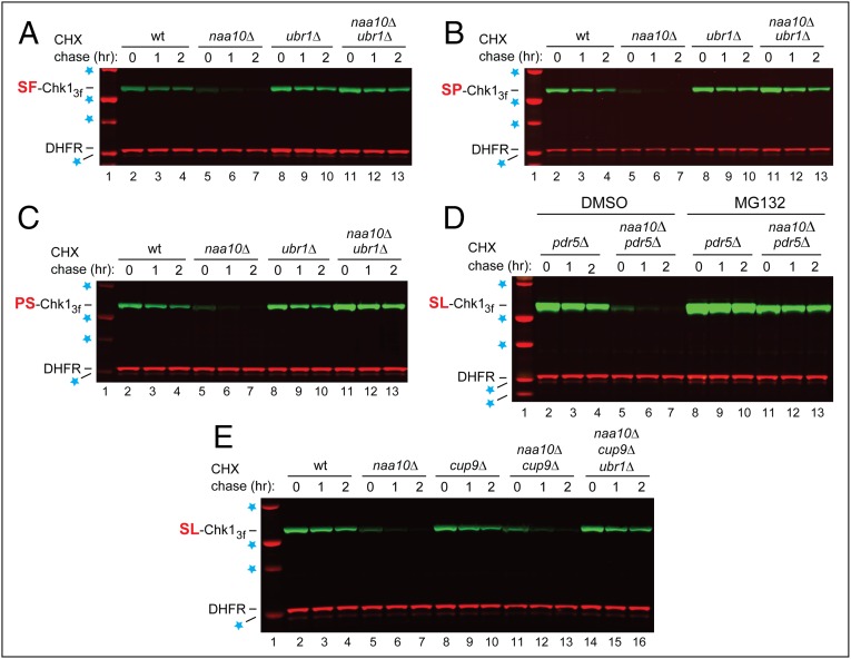
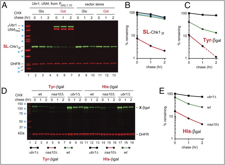
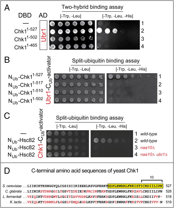
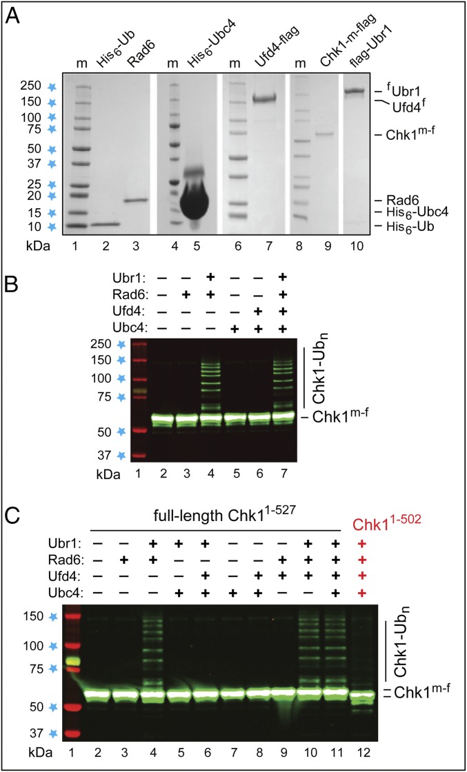
We found that the heat shock protein 90 (Hsp90) chaperone system of the yeast Saccharomyces cerevisiae is greatly impaired in naa10Δ cells, which lack the NatA Nα-terminal acetylase (Nt-acetylase) and therefore cannot N-terminally acetylate a majority of normally N-terminally acetylated proteins, including Hsp90 and most of its cochaperones. Chk1, a mitotic checkpoint kinase and a client of Hsp90, was degraded relatively slowly in wild-type cells but was rapidly destroyed in naa10Δ cells by the Arg/N-end rule pathway, which recognized a C terminus-proximal degron of Chk1. Diverse proteins (in addition to Chk1) that are shown here to be targeted for degradation by the Arg/N-end rule pathway in naa10Δ cells include Kar4, Tup1, Gpd1, Ste11, and also, remarkably, the main Hsp90 chaperone (Hsc82) itself. Protection of Chk1 by Hsp90 could be overridden not only by ablation of the NatA Nt-acetylase but also by overexpression of the Arg/N-end rule pathway in wild-type cells. Split ubiquitin-binding assays detected interactions between Hsp90 and Chk1 in wild-type cells but not in naa10Δ cells. These and related results revealed a major role of Nt-acetylation in the Hsp90-mediated protein homeostasis, a strong up-regulation of the Arg/N-end rule pathway in the absence of NatA, and showed that a number of Hsp90 clients are previously unknown substrates of the Arg/N-end rule pathway.

Keywords: Chk1; Naa10; Ubr1; Ufd4; protein degradation.


Conflict of interest statement

The authors declare no conflict of interest.

Figures

Fig. 1.
Fig. 1.
The N-end rule pathways. N-terminal residues at the top of the diagram are indicated by single-letter abbreviations. Twenty DNA-encoded amino acids are arranged to delineate three sets of N-degrons in S. cerevisiae, corresponding to three N-end rule pathways. N-terminal Met is cited twice, because it can be recognized by the Ac/N-end rule pathway (as Nt-acetylated N-terminal Met) and by the Arg/N-end rule pathway (as unacetylated N-terminal Met). N-terminal Cys is also cited twice, because it can be recognized by the Ac/N-end rule pathway (as Nt-acetylated Cys) and by the Arg/N-end rule pathway (as an oxidized, Nt-arginylatable N-terminal Cys, denoted as Cys* and formed in multicellular eukaryotes but apparently not in unstressed S. cerevisiae). (A) The Ac/N-end Rule pathway. (B) The Arg/N-end rule pathway. (C) The Pro/N-end rule pathway. See the Introduction for references and brief descriptions of these pathways.
Fig. 2.
Fig. 2.
The PRT and degradation of Chk1 in naa10Δ cells. (A and B) The PRT. See the main text for a discussion. (C) Lane 1, kDa markers. CHX-chases, using PRT with wild-type SL-Chk13f, were performed at 30 °C for 0, 20, 60, and 120 min, with wild-type (lanes 2–5), naa10Δ (lanes 6–9), ubr1Δ (lanes 10–13), and naa10Δ ubr1Δ (lanes 14–17) S. cerevisiae. Extracts were prepared from cells withdrawn at the indicated times of a chase. Proteins in an extract were fractionated by SDS/PAGE, followed by immunoblotting with anti-flag and anti-HA antibodies. (D) As in C, but CHX-chases, with the SK-Chk13f mutant, were for 0, 1, and 2 h, with wild-type (lanes 2–4), naa10Δ (lanes 5–7), ubr1Δ (lanes 8–10), and naa10Δ ubr1Δ (lanes 11–13) S. cerevisiae. (E and F) Quantification of data in C and D, respectively. For curve designations, see the keys below the immunoblots in C and D. All degradation assays in this study were performed at least twice, yielding results that differed by less than 10%. (G) As in C, but CHX-chases, with wild-type SL-Chk13f, were for 0, 1, and 2 h, with wild-type (lanes 2–4), naa10Δ (lanes 5–7), naa20Δ (lanes 8–10), and naa30Δ (lanes 11–13) S. cerevisiae. In C, D, and G, lane 1 shows kDa markers and blue stars denote 25-, 37-, 50-, and 75-kDa proteins.
Fig. 3.
Fig. 3.
Degradation of Hsc82 by the Arg/N-end rule pathway in naa10Δ cells. (A) CHX-chases, using PRT (Fig. 2B), with wild-type SL-Chk13f, for 0, 1, and 2 h, with naa10Δ S. cerevisiae carrying a high-copy plasmid that expressed Naa10 from the PGAL promoter. Lane 1, kDa markers. Blue stars denote 20-, 25-, 37-, 50-, and 75-kDa markers, respectively. Lanes 2–4 and 5–7, cells (containing vector alone) in glucose and galactose medium, respectively. Lanes 8–10 and 11–13 are as in lanes 2–4 and 5–7, respectively, but with cells containing the PGAL-NAA10 plasmid. The presence of Naa10 was verified directly, using an affinity-purified antibody to Naa10 (bands in lanes 11–13, above the bands of DHFR). (B) Immunoblotting-based comparison of the levels of endogenous (untagged) Ubr1 in wild-type naa10Δ and ubr1Δ S. cerevisiae (with ubr1Δ cells as a negative control), using an affinity-purified antibody to Ubr1 (see the main text), with immunoblots of tubulin as a loading control. Blue stars denote 50-, 75-, 100-, 150-, and 250-kDa markers, respectively. (C) Quantification of the data in B, with subtraction of the background-level signal in lane 4 (ubr1Δ cells) and with the level of Ubr1 in wild-type cells taken as 100%. (D) Degradation of Hsc82 in naa10Δ cells. Blue stars denote 25-, 37-, 50-, 75-, and 100-kDa markers, respectively. Tc-chases, using PRT (Fig. 2B) with wild-type Hsc823f, were for 0, 1, 2, and 4 h, with wild-type (lanes 2–5), naa10Δ (lanes 6–9), and ubr1Δ (lanes 10–13) S. cerevisiae. Extracts were prepared from cells withdrawn at the indicated times of a chase. Proteins in an extract were fractionated by SDS/PAGE, followed by immunoblotting with anti-flag and anti-HA antibodies. (E) Quantification of data in D. For curve designations, see the keys below the immunoblot in D. All chases in this study were performed at least twice, yielding results that differed by less than 10%.
Fig. 4.
Fig. 4.
Degradation of Chk1 mutants by the Arg/N-end rule pathway. (A) As in Fig. 2D, but with the SF-Chk13f mutant. CHX-chases, using PRT (Fig. 2B), with SF-Chk13f, for 0, 1, and 2 h, with wild-type (lanes 2–4), naa10Δ (lanes 5–7), ubr1Δ (lanes 8–10), and naa10Δ ubr1Δ (lanes 11–13) S. cerevisiae. Blue stars are as in Fig. 2D. (B) As in A but with the SP-Chk13f mutant. (C) As in A but with the PS-Chk13f mutant. (D) Lanes 2–4, CHX-chase, using PRT (Fig. 2B), with wild-type SL-Chk13f, for 0, 1, and 2 h, in pdr5Δ cells. Lanes 5–7 are as in lanes 2–4 but in pdr5Δ naa10Δ cells. Lanes 8–10 and 11–13 are as in lanes 2–4 and 5–7, respectively, but in the presence of 50 μM MG132, a proteasome inhibitor. The absence of the Pdr5 multidrug transporter was necessary to make S. cerevisiae sensitive to MG132. Blue stars denote 20-, 25-, 37-, 50-, and 75-kDa markers. (E) CHX-chases, using PRT (Fig. 2B), with wild-type SL-Chk13f, for 0, 1, and 2 h, with wild-type (lanes 2–4), naa10Δ (lanes 5–7), cup9Δ (lanes 8–10), naa10Δ cup9Δ (lanes 11–13), and naa10Δ cup9Δ ubr1Δ (lanes 14–16) S. cerevisiae. Blue stars denote 25-, 37-, 50-, and 75-kDa markers.
Fig. 5.
Fig. 5.
Overexpression of the Arg/N-end rule pathway destabilizes Chk1. (A) CHX-chases, using PRT (Fig. 2B), with wild-type SL-Chk13f, for 0, 1, and 2 h, in wild-type S. cerevisiae carrying a high-copy plasmid that expressed both flag-tagged Ubr1 (fUbr1) and Hsv-tagged Ufd4 (Ufd4hsv) from the bidirectional PGAL1,10 promoter. Lanes 2–4 and 5–7: cells containing the PGAL1,10-UBR1/UFD4 plasmid in glucose and galactose medium, respectively. Lanes 8–10 and 11–13 are as in lanes 2–4 and 5–7, respectively, but with cells containing a vector alone. The presence of fUbr1 and Ufd4hsv in lanes 5–7 was verified directly, using anti-flag and anti-Hsv antibodies. Blue stars denote 20-, 25-, 37-, 50-, 75-, 100-, 150-, and 250-kDa markers. (B) Quantification of data in A. Rhombuses, squares, triangles, and crosses denote, respectively, the decay curves of SL-Chk13f in wild-type cells overexpressing Ubr1-Ufd4 (in galactose), or containing vector alone (in glucose), or containing the repressed Ubr1-Ufd4 plasmid (in glucose), or containing vector alone (in galactose). (C) Quantification of data in D, lanes 2–10. For curve designations, see the keys below the immunoblot in D. (D) Lane 1, kDa markers. CHX-chase, using PRT (Fig. 2B), for 0, 1, and 2 h, with wild-type (lanes 2–4), naa10Δ (lanes 5–7), and ubr1Δ (lanes 8–10) S. cerevisiae expressing Tyr–β-gal, derived from Ub–Tyr–β-gal. Lanes 11–19 are as in lanes 2–10 but with His–β-gal, derived from Ub–His–β-gal. Blue stars indicate 37-, 50-, 75-, 100-, and 150-kDa markers. (E) Quantification of data in D, lanes 11–19. For curve designations, see the keys below the immunoblot in D. All chases in this study were performed at least twice, yielding results that differed by less than 10%.
Fig. 6.
Fig. 6.
Mapping Chk1 degron by two-hybrid and split-ubiquitin assays. (A) Two-hybrid binding assays with Ubr1 vs. Chk1. In both two-hybrid and split-Ub assays, the expression of HIS3 (the ultimate readout of both assays), in otherwise His cells, was a function of affinity between test proteins. (AC, Left) Images of His-containing plates, on which all yeast strains grew. (Right) His-lacking plates, on which only His+ cells grew. A two-hybrid-based Ubr1 fusion bearing the activation domain (AD) was examined vs. full-length Chk11–527 and its truncated derivatives bearing the DNA-binding domain (DBD). (A, 1) Ubr1 alone (negative control). (A, 2) Full-length Chk11–527 vs. Ubr1. (A, 3) Chk11–502 vs. Ubr1. (A, 4) Chk11–465 vs. Ubr1. (B) Split-Ub binding assays with Ubr1 vs. Chk1. Split-Ub–based fusions (Materials and Methods) of Ubr1 vs. Chk11–527, Chk11–517, Chk11–510, and Chk11–502. (C) Split-Ub binding assays with full-length Chk1 vs. full-length Hsc82 in wild-type, naa10Δ, and naa10Δ ubr1Δ cells. C1, Chk1 alone (negative control). (D) Alignment of C-terminal regions of Chk1 from the indicated yeast species. The last 25 residues of S. cerevisiae Chk1 are highlighted in yellow. The last 10 residues of Chk1 (found to be essential for the binding to Ubr1; see C, 2) are marked as well. Residues of Chk1 in species other than S. cerevisiae that differed from those of S. cerevisiae Chk1 are in red.
Fig. 7.
Fig. 7.
Degron-specific polyubiquitylation of Chk1 in a defined in vitro system. (A) Coomassie-stained SDS/PAGE patterns of purified S. cerevisiae His6-Ub, Rad6, His6-Ubc4, Ufd4f, full-length SL-Chk1mf1527, and fUbr1. (B) Lane 1, kDa markers. Lanes 2–7, SL-Chk1mf1527 incubated in the presence of ATP, Ub, Uba1, and other purified proteins as indicated. Immunoblotting with anti-flag antibody. (C) As in B but independent Chk1 polyubiquitylation assays that included, in addition, the assay with C-terminally truncated SL-Chk1mf1502 (lane 12).

Similar articles

Cited by

References

    1. Hartl FU, Bracher A, Hayer-Hartl M. Molecular chaperones in protein folding and homeostasis. Nature. 2011;475:324–332. - PubMed
    1. Horwich AL. Molecular chaperones in cellular protein folding: The birth of a field. Cell. 2014;157:285–288. - PubMed
    1. Wolff S, Weissman JS, Dillin A. Differential scales of protein quality control. Cell. 2014;157:52–64. - PubMed
    1. Powers ET, Balch WE. Diversity in the origins of proteostasis networks––a driver for protein function in evolution. Nat Rev Mol Cell Biol. 2013;14:237–248. - PMC - PubMed
    1. Borkovich KA, Farrelly FW, Finkelstein DB, Taulien J, Lindquist S. hsp82 is an essential protein that is required in higher concentrations for growth of cells at higher temperatures. Mol Cell Biol. 1989;9:3919–3930. - PMC - PubMed

Publication types

MeSH terms