. 2017 Jun 23;292(25):10321-10327.
doi: 10.1074/jbc.C117.790311. Epub 2017 May 17.

The C-terminal peptide of Aquifex aeolicus riboflavin synthase directs encapsulation of native and foreign guests by a cage-forming lumazine synthase

Affiliations

The C-terminal peptide of Aquifex aeolicus riboflavin synthase directs encapsulation of native and foreign guests by a cage-forming lumazine synthase

Yusuke Azuma et al. J Biol Chem. .

Abstract

Encapsulation of specific enzymes in self-assembling protein cages is a hallmark of bacterial compartments that function as counterparts to eukaryotic organelles. The cage-forming enzyme lumazine synthase (LS) from Bacillus subtilis (BsLS), for example, encapsulates riboflavin synthase (BsRS), enabling channeling of lumazine from the site of its generation to the site of its conversion to vitamin B2 Elucidating the molecular mechanisms underlying the assembly of these supramolecular complexes could help inform new approaches for metabolic engineering, nanotechnology, and drug delivery. To that end, we investigated a thermostable LS from Aquifex aeolicus (AaLS) and found that it also forms cage complexes with the cognate riboflavin synthase (AaRS) when both proteins are co-produced in the cytosol of Escherichia coli A 12-amino acid-long peptide at the C terminus of AaRS serves as a specific localization sequence responsible for targeting the guest to the protein compartment. Sequence comparisons suggested that analogous peptide segments likely direct RS complexation by LS cages in other bacterial species. Covalent fusion of this peptide tag to heterologous guest molecules led to their internalization into AaLS assemblies both in vivo and in vitro, providing a firm foundation for creating tailored biomimetic nanocompartments for medical and biotechnological applications.

Keywords: bacterial compartment; biotechnology; cell compartmentalization; localization sequence; lumazine/riboflavin synthase; protein cage; protein self-assembly; protein-protein interaction; synthetic biology.


Conflict of interest statement

The authors declare that they have no conflicts of interest with the contents of this article

Figures
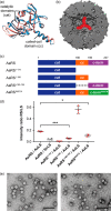
Figure 1.
Figure 1.
Complexation of AaRS by AaLS in E. coli cells. a, a homology model of the AaRS monomer (blue) superimposed on the riboflavin synthase from S. pombe (red). The model was taken from the ModWeb server for protein modeling (37). The model ID is d5f76bef49077cd7ae8f8c7269bb2ad5. The catalytic domains (cat) and the coiled-coil domain (cc) are indicated. b, cutaway view along one of the 3-fold symmetry axes of the AaLS cage (PDB ID: 1HQK) (19). The clefts constituting the enzyme catalytic site are highlighted in red. c, genes encoding AaRS and engineered constructs. The structurally disordered C-terminal peptide is designated as C-term. d, intensity ratio of riboflavin synthase/lumazine synthase determined by SDS-PAGE analysis. *** and * signify p < 0.001 and 0.1, respectively; n.d., not detected. Error bars indicate standard deviations from triplicate experiments. e, TEM images of empty AaLS assemblies (left) and complexes with AaRS (right). Scale bar = 100 nm.
Figure 2.
Figure 2.
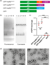
Loading a foreign guest protein into AaLS cages using a C-terminal peptide from AaRS. a, genes encoding GFP fused to the sequence encoding the C terminus of AaRS. C-term and cc indicate the structurally disordered C-terminal peptide and coiled-coil domain, respectively. C-termscr is a scrambled version of AaRS196–207. b, native AGE analysis of co-expressed and purified GFP·AaLS. Lane 1, GFP-AaRS180–207/AaLS; lane 2, GFP-AaRS196–207/AaLS; lane 3, GFP-AaRS180–207scr/AaLS; lane 4, GFP·AaLS; lane 5, AaLS; lane 6, His-GFP. c, estimated numbers of GFP molecules per AaLS cage as determined from UV-visible absorbance spectra. ***, p < 0.001. ns, not significant. Error bars indicate standard deviation from triplicate experiments. d, transmission electron microscopy images of AaLS assemblies complexed with GFP-AaRS180–207 (left) and GFP-AaRS196–207 (right). The particles closely resemble the wild-type assemblies shown in Fig. 1e. Scale bar = 100 nm.
Figure 3.
Figure 3.
Characterization of the AaRS C-terminal dodecapeptide in vitro. a, peptide sequences. Fl = FITC-labeled N terminus, J = aminocaproic acid. b, CD spectra of AaRS196–207 (solid black line) and AaRS196–207scr (dotted gray line) in aqueous buffer (left) and 30% trifluoroethanol (right). deg, degree. c, hydrodynamic size of AaLS assemblies as a function of temperature (open squares) or GdnHCl concentration (filled circles). Crystallographic data show that AaLS icosahedral assemblies and AaLS pentamers have diameters of ∼16 and ∼9 nm, respectively (19). These values are indicated by gray dashed lines. d, TEM image of AaLS after treatment with 3 m GdnHCl. Scale bar = 100 nm. e, AGE analysis of in vitro assembled complexes between fluorescently labeled AaRS peptides and AaLS. f, TEM image of Fl-AaRS196–207/AaLS complexes obtained after GdnHCl treatment. Scale bar = 100 nm. g, loading efficiency of the FITC-labeled peptide into AaLS, determined by absorbance spectra. Error bars indicate standard deviation from duplicate experiments.

References

    1. Bobik T. A. (2006) Polyhedral organelles compartmenting bacterial metabolic processes. Appl. Microbiol. Biotechnol. 70, 517–525 - PubMed
    1. Yeates T. O., Kerfeld C. A., Heinhorst S., Cannon G. C., and Shively J. M. (2008) Protein-based organelles in bacteria: carboxysomes and related microcompartments. Nat. Rev. Microbiol. 6, 681–691 - PubMed
    1. Kerfeld C. A., Heinhorst S., and Cannon G. C. (2010) Bacterial microcompartments. Annu. Rev. Microbiol. 64, 391–408 - PubMed
    1. Sutter M., Boehringer D., Gutmann S., Günther S., Prangishvili D., Loessner M. J., Stetter K. O., Weber-Ban E., and Ban N. (2008) Structural basis of enzyme encapsulation into a bacterial nanocompartment. Nat. Struct. Mol. Biol. 15, 939–947 - PubMed
    1. Fan C., Cheng S., Liu Y., Escobar C. M., Crowley C. S., Jefferson R. E., Yeates T. O., and Bobik T. A. (2010) Short N-terminal sequences package proteins into bacterial microcompartments. Proc. Natl. Acad. Sci. U.S.A. 107, 7509–7514 - PMC - PubMed

MeSH terms

LinkOut - more resources