. 2017 Jun 30;292(26):10865-10882.
doi: 10.1074/jbc.M117.788950. Epub 2017 May 17.

The h-region of twin-arginine signal peptides supports productive binding of bacterial Tat precursor proteins to the TatBC receptor complex

Affiliations

The h-region of twin-arginine signal peptides supports productive binding of bacterial Tat precursor proteins to the TatBC receptor complex

Agnes Ulfig et al. J Biol Chem. .

Abstract

The twin-arginine translocation (Tat) pathway transports folded proteins across bacterial membranes. Tat precursor proteins possess a conserved twin-arginine (RR) motif in their signal peptides that is involved in their binding to the Tat translocase, but some facets of this interaction remain unclear. Here, we investigated the role of the hydrophobic (h-) region of the Escherichia coli trimethylamine N-oxide reductase (TorA) signal peptide in TatBC receptor binding in vivo and in vitro We show that besides the RR motif, a minimal, functional h-region in the signal peptide is required for Tat-dependent export in Escherichia coli Furthermore, we identified mutations in the h-region that synergistically suppressed the export defect of a TorA[KQ]-30aa-MalE Tat reporter protein in which the RR motif was replaced with a lysine-glutamine pair. Strikingly, all suppressor mutations increased the hydrophobicity of the h-region. By systematically replacing a neutral residue in the h-region with various amino acids, we detected a positive correlation between the hydrophobicity of the h-region and the translocation efficiency of the resulting reporter variants. In vitro cross-linking of residues located in the periplasmically-oriented part of the TatBC receptor to TorA[KQ]-30aa-MalE reporter variants harboring a more hydrophobic h-region in their signal peptides confirmed that unlike in TorA[KQ]-30aa-MalE with an unaltered h-region, the mutated reporters moved deep into the TatBC-binding cavity. Our results clearly indicate that, besides the Tat motif, the h-region of the Tat signal peptides is another important binding determinant that significantly contributes to the productive interaction of Tat precursor proteins with the TatBC receptor complex.

Keywords: Escherichia coli (E. coli); membrane transport; protein export; protein targeting; protein translocation.


Conflict of interest statement

The authors declare that they have no conflicts of interest with the contents of this article.

Figures

Figure 1.
Figure 1.
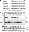
Effect of the V23D mutation in the h-region of the TorA signal peptide on the export of TorA-MalE. A, amino acid sequence of TorA-MalE encompassing the Tat consensus motif (dashed line) and the entire h-region of the TorA signal peptide. The position of the introduced V23D mutation is marked by an arrow. B, subcellular localization of TorA-MalE-derived polypeptides. Cells were fractionated into a periplasmic (P) fraction and a combined C/M fraction by EDTA-lysozyme spheroplasting. The samples of the fractions corresponding to an identical amount of cells were subjected to SDS-PAGE and immunoblotting using anti-MalE antibodies. The positive control was E. coli GSJ101 containing plasmids pTorA-MalE and pHSG-TatABCE (lanes 3 and 4). The other samples correspond to GSJ101 containing plasmids pTorA[V23D]-MalE and pHSG-TatABCE (lanes 1 and 2) and GSJ101 containing pTorA-MalE and the pHSG575 empty vector (lanes 5 and 6). wt, wild-type; p, TorA-MalE/TorA[V23D]-MalE precursor in the C/M fraction; m, mature form of MalE in the P fraction; asterisk, TorA-MalE/TorA[V23D]-MalE-derived degradation products in the C/M fraction. Positions of molecular weight markers are indicated on the left margin. The phenotypes of the respective strains on MMM (−, no growth; +++: fast growth) and MCM (pale, red) agar plates are shown in the boxes at the bottom of the figure.
Figure 2.
Figure 2.
Export of the TorA[V23D]-MalE precursor protein is restored by h-region-located suppressor mutations. A, amino acid sequences of TorA-MalE reporter variants encompassing the Tat consensus motif (dashed line) and the entire h-region of the TorA signal peptide. Positions of the particular mutations are underlined. Deletions are visualized as gaps. B, subcellular localization of TorA-MalE-derived polypeptides. Cells were fractionated into a periplasmic (P) fraction and a combined C/M fraction by EDTA-lysozyme spheroplasting. The samples of the fractions corresponding to an identical amount of cells were subjected to SDS-PAGE and immunoblotting using anti-MalE antibodies. The positive control was E. coli GSJ101 containing plasmids pTorA-MalE and pHSG-TatABCE (lane 1). The other samples correspond to GSJ101 co-expressing the tatABCE genes and no reporter protein (none; lane 7), the export-defective TorA[V23D]-MalE reporter (lane 2), or one of the isolated reporter variants containing the mutations indicated above the lanes (lanes 3–6). wt, wild-type; p, wt or mutant TorA-MalE precursor in the C/M fraction; m, mature form of MalE in the P fraction; asterisk, wt or mutant TorA-MalE-derived degradation products in the C/M fraction. Positions of molecular mass markers are indicated on the left margin. The phenotypes of the respective strains on MMM (−, no growth; ++, moderate growth; +++, fast growth) and MCM (pale, pink, red) agar plates in the presence (Tat+) or absence (Tat) of the Tat translocase are shown in the boxes at the bottom of the figure.
Figure 3.
Figure 3.
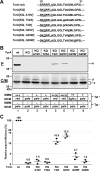
Suppression of the export-defect of a TorA[KQ]-30aa-MalE precursor protein by mutations in the h-region of the TorA signal peptide. A, amino acid sequences of TorA-30aa-MalE reporter variants encompassing the Tat consensus motif (dashed line) and the entire h-region of the TorA signal peptide. Positions of the particular mutations are underlined. B, subcellular localization of TorA-30aa-MalE-derived polypeptides. Cells were fractionated into a P fraction and a combined C/M fraction by EDTA-lysozyme spheroplasting. The samples of the fractions corresponding to an identical amount of cells were subjected to SDS-PAGE and immunoblotting using anti-MalE antibodies. The positive control was E. coli GSJ101 containing plasmids pTorA-30aa-MalE and pHSG-TatABCE (lane 1). The other samples correspond to GSJ101 co-expressing the tatABCE genes and the export-defective TorA[KQ]-30aa-MalE reporter (lane 2) or one of the isolated reporter variants containing the single mutations in the h-region indicated above the lanes (lanes 3–8). wt, wild-type; p, wt, or mutated TorA-30aa-MalE precursor in the C/M fraction; m, mature form of MalE in the P fraction; asterisk, wt, or mutated TorA-30aa-MalE-derived degradation products in the C/M fraction. Positions of molecular weight markers are indicated on the left margin. The phenotypes of the respective strains on MMM (−, no growth; +, slow growth; ++, moderate growth; +++, fast growth) and MCM (pale, light red, red) agar plates in the presence (Tat+) or absence (Tat) of the Tat translocase are shown in the boxes at the bottom of the figure. C, relative export efficiencies of the analyzed TorA-30aa-MalE reporter variants in strains expressing the Tat translocase. The amount of exported MalE protein in the P fraction of strains GSJ101 co-expressing the tatABCE genes and TorA-30aa-MalE or one of the mutated TorA[KQ]-30aa-MalE reporter variants was determined in at least three different independent experiments via quantification of the chemiluminescence signals. The signals were recorded by a CCD camera and subsequently analyzed by the program AIDA 4.50 (Raytest). The average values are indicated by horizontal marker lines; standard deviations by error bars. The relative export efficiency of the positive control GSJ101 (pTorA-30aa-MalE, pHSG-TatABCE) was set to 100%.
Figure 4.
Figure 4.
Combinations of mutations in the h-region of the TorA signal peptide synergistically suppress the export-defect of a TorA[KQ]-30aa-MalE precursor protein. A, amino acid sequences of TorA-30aa-MalE reporter variants encompassing the Tat consensus motif (dashed line) and the entire h-region of the TorA signal peptide. Positions of the particular mutations are underlined. B–D, subcellular localization of TorA-30aa-MalE-derived polypeptides. Cells were fractionated into a P fraction and a combined C/M fraction by EDTA-lysozyme spheroplasting. The samples of the fractions corresponding to an identical amount of cells were subjected to SDS-PAGE and immunoblotting using anti-MalE antibodies. The positive control was E. coli GSJ101 containing plasmids pTorA-30aa-MalE and pHSG-TatABCE (lane 1). The other samples correspond to GSJ101 co-expressing the tatABCE genes and the export-defective TorA[KQ]-30aa-MalE reporter (lane 2) or one of the TorA[KQ]-30aa-MalE reporter variants containing single or double mutations in the h-region of the TorA signal peptide indicated above the lanes. wt, wild-type; p, wt or mutated TorA-30aa-MalE precursor in the C/M fraction; m, mature form of MalE in the P fraction; asterisk, wt or mutated TorA-30aa-MalE-derived degradation products in the C/M fraction. Positions of molecular mass markers are indicated on the left margin. The phenotypes of the respective strains on MMM (−, no growth; +, slow growth; ++, moderate growth; +++, fast growth) and MCM (pale, light red, red) agar plates in the presence (Tat+) or absence (Tat) of the Tat translocase are shown in the boxes at the bottom of the figure. E, relative export efficiencies of the analyzed TorA-30aa-MalE reporter variants in strains expressing the Tat translocase. The amount of exported MalE protein in the P fraction of strains GSJ101 co-expressing the tatABCE genes and TorA-30aa-MalE or one of the mutated TorA[KQ]-30aa-MalE reporter variants was determined in at least three different independent experiments via quantification of the chemiluminescence signals. The signals were recorded by a CCD camera and subsequently analyzed by the program AIDA 4.50 (Raytest). The average values are indicated by horizontal marker lines; standard deviations by error bars. The relative export efficiency of the positive control GSJ101 (pTorA-30aa-MalE, pHSG-TatABCE) was set to 100%.
Figure 5.
Figure 5.
Mutations in the h-region of the TorA signal peptide suppress the export defect of a TorA[KQ]-30aa-MalE precursor protein by increasing the overall hydrophobicity of the signal peptide. A, amino acid sequences of TorA-30aa-MalE reporter variants encompassing the Tat consensus motif (dashed line) and the entire h-region of the TorA signal peptide. Positions of the particular mutations are underlined. B and C, subcellular localization of TorA-30aa-MalE-derived polypeptides. Cells were fractionated into a P fraction and a combined C/M fraction by EDTA-lysozyme spheroplasting. The samples of the fractions corresponding to an identical amount of cells were subjected to SDS-PAGE and immunoblotting using anti-MalE antibodies. The positive control was E. coli GSJ101 containing plasmids pTorA-30aa-MalE and pHSG-TatABCE (lane 1). The other samples correspond to GSJ101 co-expressing the tatABCE genes and the export-defective TorA[KQ]-30aa-MalE reporter (lane 2) or one of the TorA[KQ]-30aa-MalE reporter variants containing the single mutations in the h-region of the TorA signal peptide indicated above the lanes. wt, wild-type; p, wt or mutated TorA-30aa-MalE precursor in the C/M fraction; m, mature form of MalE in the P fraction; asterisk, wt or mutated TorA-30aa-MalE-derived degradation products in the C/M fraction. Positions of molecular mass markers are indicated on the left margin. The phenotypes of the respective strains on MMM (−, no growth; ++, moderate growth; +++, fast growth) and MCM (pale, light red, red) agar plates in the presence (Tat+) or absence (Tat) of the Tat translocase are shown in the boxes at the bottom of the figure. D, relative export efficiencies of the analyzed TorA-30aa-MalE reporter variants in strains expressing the Tat translocase. The amount of exported MalE protein in the P fraction of strains GSJ101 co-expressing the tatABCE genes and TorA-30aa-MalE or one of the mutated TorA[KQ]-30aa-MalE reporter variants was determined in at least three different independent experiments via quantification of the chemiluminescence signals. The signals were recorded by a CCD camera and subsequently analyzed by the program AIDA 4.50 (Raytest). The average values are indicated by horizontal marker lines; standard deviations by error bars. The relative export efficiency of the positive control GSJ101 (pTorA-30aa-MalE, pHSG-TatABCE) was set to 100%.
Figure 6.
Figure 6.
Suppressors of the TorA[KQ]-30aa-MalE export defect harboring mutations in the h-region of the TorA signal peptide are inserted deep into the TatBC-binding cavity. TorA-30aa-MalE reporter variants containing wild-type (wt) or mutated TorA* signal peptides harboring mutations in the RR motif and/or the h-region of the TorA signal peptide indicated above the lines were synthesized and radioactively labeled by in vitro transcription/translation in the presence of inverted E. coli INV, which contained TatABC. Bpa was incorporated either (A, C) at the Ile-4 position in the N terminus of TatB or (B) at the Val-202 position in TM5 of TatC. In samples labeled with a +, cross-linking was initiated by irradiation with ultraviolet light or mock treated (−). Radiolabeled cross-linking products were separated by SDS-PAGE and visualized by phosphorimaging. Indicated are the positions of molecular size standard proteins, the various TorA*-30aa-MalE precursor proteins (p) and the cross-linked TatB-TorA*-30aa-MalE (black star) or TatC-TorA*-30aa-MalE (black square) complexes. C, effect of CCCP and DCCD on cross-linking between residue Ile-4 in TatB and TorA[KQ, T22I]-30aa-MalE (iKQ-T22I).
Figure 7.
Figure 7.
Combinations of mutations in the h-region of the TorA signal peptide and the mutation L9F in TatC synergistically suppress the export defect of a TorA[KQ]-30aa-MalE precursor protein. A and B, subcellular localization of TorA-30aa-MalE-derived polypeptides. Cells were fractionated into a P fraction and a combined C/M fraction by EDTA-lysozyme spheroplasting. The samples of the fractions corresponding to an identical amount of cells were subjected to SDS-PAGE and immunoblotting using anti-MalE antibodies. The positive controls were E. coli GSJ101 containing plasmids pTorA-30aa-MalE and pHSG-TatABCE (lane 1) or pHSG-TatABC[L9F]E (lane 2). The other samples correspond to GSJ101 co-expressing the wild-type tatABCE or mutated tatABC[L9F]E genes and the export-defective TorA[KQ]-30aa-MalE reporter (lanes 3 and 4, respectively) or one of the TorA[KQ]-30aa-MalE reporter variants containing single mutations or double mutations in the h-region of the TorA signal peptide as indicated above the lanes. wt, wild-type; p, wt or mutated TorA-30aa-MalE precursor in the C/M fraction; m, mature form of MalE in the P fraction; asterisk, wt or mutated TorA-30aa-MalE-derived degradation products in the C/M fraction. Positions of molecular mass markers are indicated on the left margin. The phenotypes of the respective strains on MMM (−, no growth; +, slow growth; ++, moderate growth; +++, fast growth) and MCM (pale, light red, pink, red) agar plates after 15 and 24 h of incubation are shown in the boxes at the bottom of the figure. C, relative export efficiencies of the analyzed TorA-30aa-MalE reporter variants in strains expressing the wild-type or mutant TatABC[L9F]E translocase. The amount of exported MalE protein in the P fraction of strains GSJ101 co-expressing the wild-type tatABCE or mutated tatABC[L9F]E genes and TorA-30aa-MalE or one of the mutated TorA[KQ]-30aa-MalE reporter variants was determined in at least three different independent experiments via quantification of the chemiluminescence signals. The signals were recorded by a CCD camera and subsequently analyzed by the program AIDA 4.50 (Raytest). The average values are indicated by horizontal marker lines; standard deviations by error bars. The relative export efficiency of the positive control GSJ101 (pTorA-30aa-MalE, pHSG-TatABCE) was set to 100%.

Similar articles

Cited by

References

    1. Denks K., Vogt A., Sachelaru I., Petriman N. A., Kudva R., and Koch H. G. (2014) The Sec translocon mediated protein transport in prokaryotes and eukaryotes. Mol. Membr. Biol. 31, 58–84 - PubMed
    1. Robinson C., Matos C. F., Beck D., Ren C., Lawrence J., Vasisht N., and Mendel S. (2011) Transport and proofreading of proteins by the twin-arginine translocation (Tat) system in bacteria. Biochim. Biophys. Acta 1808, 876–884 - PubMed
    1. Hou B., and Brüser T. (2011) The Tat-dependent protein translocation pathway. Biomol. Concepts 2, 507–523 - PubMed
    1. Fröbel J., Rose P., and Müller M. (2012) Twin-arginine-dependent translocation of folded proteins. Philos. Trans. R. Soc. Lond. B Biol. Sci. 367, 1029–1046 - PMC - PubMed
    1. Palmer T., and Berks B. C. (2012) The twin-arginine translocation (Tat) protein export pathway. Nat. Rev. Microbiol. 10, 483–496 - PubMed

MeSH terms

LinkOut - more resources