Comparative Study
. 2018 Jan 10;17(1):28-34.
doi: 10.2463/mrms.mp.2016-0128. Epub 2017 May 18.

Evaluating Myometrial Invasion in Endometrial Cancer: Comparison of Reduced Field-of-view Diffusion-weighted Imaging and Dynamic Contrast-enhanced MR Imaging

Affiliations
Comparative Study

Evaluating Myometrial Invasion in Endometrial Cancer: Comparison of Reduced Field-of-view Diffusion-weighted Imaging and Dynamic Contrast-enhanced MR Imaging

Mayumi Takeuchi et al. Magn Reson Med Sci. .

Abstract

Purpose: To compare the diagnostic ability of reduced FOV diffusion-weighted (DW) MR imaging with that of 3D dynamic contrast-enhanced (DCE) MR imaging in evaluating the depth of myometrial invasion in patients with endometrial cancer.

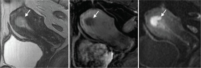
Methods: Three tesla MR images including T2-weighted imaging, reduced FOV DW imaging and 3D DCE MR imaging in sagittal and oblique axial (short axis) planes in 25 women with surgically proven endometrial cancer were retrospectively evaluated. The depth of myometrial invasion (stage S: < 50% vs stage D: ≥ 50%) on MR imaging was correlated with surgical pathology results.

Results: The 25 endometrial cancers included 16 stage S and 9 stage D tumors. The depth of myometrial invasion could be accurately evaluated in 68% of the cases for T2-weighted imaging, 92% for 3D DCE MR imaging, and 96% for reduced FOV DW imaging. In two patients with coexisting adenomyosis, both T2-weighted imaging and 3D DCE MR imaging failed to reveal the deep myometrial invasion, and reduced FOV DW imaging clearly demonstrated the tumor margin in the cases. Combination of reduced FOV DW imaging reading together with T2-weighted imaging improved the assessment of myometrial invasion with a diagnostic accuracy of up to 100%.

Conclusions: Addition of reduced FOV DW imaging may improve the staging accuracy of MR imaging for endometrial cancer in assessing the depth of myometrial invasion. Especially, reduced FOV DW imaging has an advantage in assessing the depth of myometrial invasion for patients with coexisting adenomyosis. Reduced FOV DW imaging can be an alternative to 3D DCE MR imaging in evaluating myometrial invasion of endometrial cancer without the use of contrast medium.

Keywords: diffusion-weighted magnetic resonance imaging; dynamic contrast-enhanced-magnetic resonance imaging; endometrial cancer; magnetic resonance imaging; reduced field-of-view.


Conflict of interest statement

Conflicts of Interest

The authors declare that they have no conflicts of interest.

Figures

Fig. 1.
Fig. 1.
A 54-year-old woman with endometrial cancer (stage S). (a) Sagittal fast spin-echo T2-weighted image shows intermediate signal intensity endometrial mass (arrow) within the endometrium. No deep myometrial invasion is revealed, however, the boundary between the tumor and the endometrium is not clear due to poor contrast. (b) Sagittal contrast-enhanced T1-weighted imaging shows the tumor (arrow) as low intensity lesion, which contrasts well with well-enhanced myometrium without deep myometrial invasion. (c) Sagittal reduced FOV Diffusion-weighted (DW) imaging shows high signal intensity endometrial mass (arrow), which contrasts well with both intermediate signal intensity endometrium and low signal intensity myometrium without deep myometrial invasion.
Fig. 2.
Fig. 2.
A 72-year-old woman with endometrial cancer (stage D). (a) Oblique axial fast spin-echo T2-weighted image with fat saturation shows intermediate signal intensity large endometrial mass (arrow). The boundary between the tumor and the myometrium is not clear due to poor contrast, and assessment of the depth of myometrial invasion is not possible. (b) Oblique axial contrast-enhanced T1-weighted imaging shows the tumor (arrow) as low intensity lesion, which contrasts well with well-enhanced myometrium with deep myometrial invasion. (c) Oblique axial reduced FOV Diffusion-weighted (DW) imaging shows high signal intensity endometrial mass (arrow), which contrasts well with low signal intensity myometrium with deep myometrial invasion.
Fig. 3.
Fig. 3.
A 59-year-old woman with endometrial cancer (stage D). (a) Oblique axial fast spin-echo T2-weighted image with fat saturation shows intermediate signal intensity endometrial mass (arrow). The boundary between the tumor and the myometrium is clear, however, the tumor margin adjacent to coexisting adenomyosis is unclear due to poor contrast, and assessment of the depth of myometrial invasion is difficult. (b) On oblique axial contrast-enhanced T1-weighted imaging, the boundary between low intensity tumor (arrow) and heterogeneously enhanced myometrium with coexisting adenomyosis is not clear due to poor contrast, and assessment of the depth of myometrial invasion is not possible. (c) Oblique axial reduced FOV Diffusion-weighted (DW) imaging shows high signal intensity endometrial mass with deep myometrial invasion (arrow), which contrasts well with low signal intensity myometrium with coexisting adenomyosis.
Fig. 4.
Fig. 4.
A 59-year-old woman with endometrial cancer (stage S). (a) Oblique axial fast spin-echo T2-weighted image with fat saturation shows intermediate signal intensity endometrial mass (arrow). The boundary between the tumor and the myometrium is clear with preserved low signal intensity junctional zone. (b) Oblique axial contrast-enhanced T1-weighted imaging shows the tumor (arrow) as low intensity mass, which contrasts well with well-enhanced myometrium without deep myometrial invasion. (c) Oblique axial reduced FOV Diffusion-weighted (DW) imaging shows high signal intensity endometrial mass (arrow). Assessment of the depth of myometrial invasion is not possible due to distortion caused by susceptibility artifact from adjacent rectal gas.

Similar articles

Cited by

References

    1. Amant F, Mirza MR, Creutzberg CL. Cancer of the corpus uteri. Int J Gynaecol Obstet 2012; 119 Suppl 2:S110–S117. - PubMed
    1. Ludwig H. Prognostic factors in endometrial cancer. Int J Gynecol Obstet 1995; 49:S1–S7. - PubMed
    1. Larson DM, Connor GP, Broste SK, Krawisz BR, Johnson KK. Prognostic significance of gross myometrial invasion with endometrial cancer. Obstet Gynecol 1996; 88:394–398. - PubMed
    1. Frei KA, Kinkel K. Staging endometrial cancer: role of magnetic resonance imaging. J Magn Reson Imaging 2001; 13:850–855. - PubMed
    1. Beddy P, Moyle P, Kataoka M, et al. Evaluation of depth of myometrial invasion and overall staging in endometrial cancer: comparison of diffusion-weighted and dynamic contrast-enhanced MR imaging. Radiology 2012; 262:530–537. - PubMed

Publication types

MeSH terms