. 2017 May 17;7(1):2022.
doi: 10.1038/s41598-017-02222-0.

Protein kinase C inhibitor chelerythrine selectively inhibits proliferation of triple-negative breast cancer cells

Affiliations

Protein kinase C inhibitor chelerythrine selectively inhibits proliferation of triple-negative breast cancer cells

Wanjun Lin et al. Sci Rep. .

Abstract

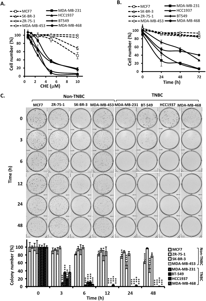
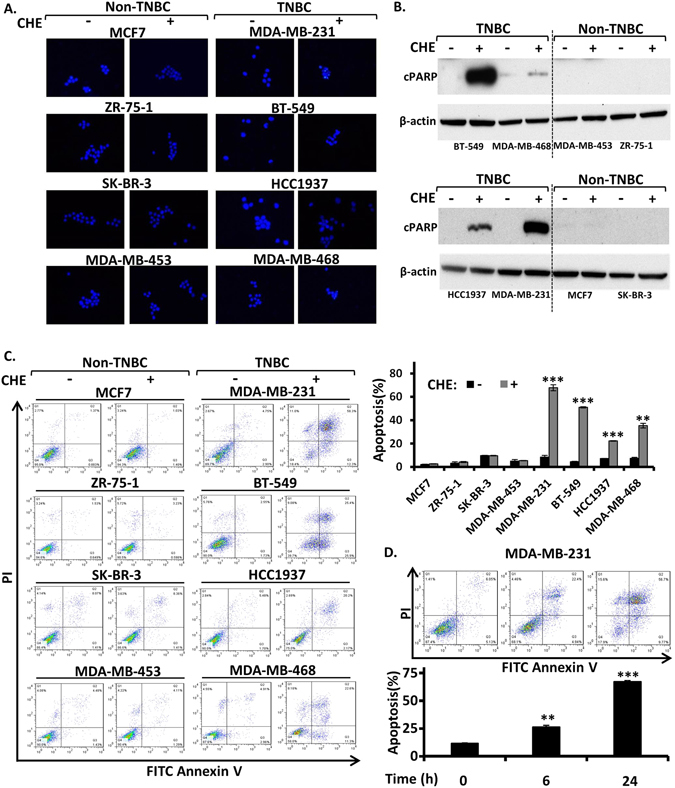
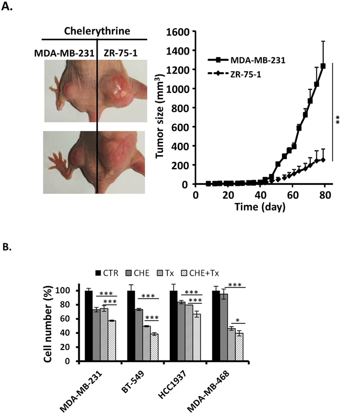
Triple-negative breast cancer (TNBC) is a subtype of breast cancer lacking targeted therapy currently. Recent studies imply that protein kinase C may play important roles in TNBC development and could be a specific target. In this study, we evaluated the anti-proliferative activity of PKC inhibitor chelerythrine on a panel of breast cancer cell lines. Chelerythrine selectively inhibited the growth of TNBC cell lines compared to non-TNBC cell lines as demonstrated by in vitro cell proliferation assay and colony formation assay, as well as evidenced by in vivo xenograft assay. The selective anti-proliferative effect of chelerythrine was associated with induction of apoptosis in TNBC cell lines. We further demonstrated that PKN2, one of the PKC subtypes, was highly expressed in TNBC cell lines, and knocking down PKN2 in TNBC cells inhibited colony formation and xenograft growth. This indicates that PKN2 is required for the survival of TNBC cells, and could be the target mediates the selective activity of chelerythrine. Finally, combination of chelerythrine and chemotherapy reagent taxol showed synergistic/additive effect on TNBC cell lines. Our results suggest chelerythrine or other PKC inhibitors may be promising regimens for TNBC tumors.


Conflict of interest statement

The authors declare that they have no competing interests.

Figures

Figure 1
Figure 1
Chelerythrine selectively inhibits proliferation of TNBC cell lines in vitro. Four non-triple-negative breast cancer (Non-TNBC) cell lines (MCF7, ZR-75-1, SK-BR-3, MDA-MB-453) and four triple-negative breast cancer (TNBC) cell lines (MDA-MB-231, BT549, HCC1937 and MDA-MB-468) were used to determine the growth inhibition effect of chelerythrine (CHE). (A) Dose effect of chelerythrine treatment (72 hours) on the proliferation of non-TNBC cell lines compared with TNBC cell lines. The cell number at each chelerythrine concentration is represented as a percentage of control (no chelerythrine treatment). Average values are from three independent experiments performed in duplicate (n = 3). (B) Time course of chelerythrine treatment (5 μM) on the proliferation of non-TNBC cell lines compared with TNBC cell lines. The cell number at each time point is represented as a percentage of control (no chelerythrine treatment). Average values are from three independent experiments performed in duplicate (n = 3). (C) Colony formation after treatment with chelerythrine (5 μM) for indicated times. Representative colony formation assay plates are shown, which were quantified by counting colony number (n = 4). Data are shown as mean ± SD. P-values determined by Student’s t-test compared to control. *P < 0.05; **P < 0.01; ***P < 0.001.
Figure 2
Figure 2
Chelerythrine selectively induces apoptosis in TNBC cell lines. Four non-triple-negative breast cancer (Non-TNBC) cell lines (MCF7, ZR-75-1, SK-BR-3, MDA-MB-453) and four triple-negative breast cancer (TNBC) cell lines (MDA-MB-231, BT549, HCC1937 and MDA-MB-468) were treated with chelerythrine (CHE, 5 μM) for 24 hours. (A) Visualization of apoptotic morphological changes by fluorescent microscope with Hoechst 33258 staining. Reprehensive pictures are shown (400x). (B) Western blotting analysis of apoptosis marker cleaved nuclear poly (ADP-ribose) polymerase (cPARP). (C) Representative contour diagrams of FITC Annexin V/PI flow cytometry analysis of cells. Fractions of apoptotic cells were quantified. Average values are from three independent experiments (n = 3). (D) TNBC cell line MDA-MB-231 was treated with chelerythrine (CHE, 5 μM) for 0, 6 and 24 hours. Apoptosis was analyzed by flow cytometry with Annexin V/PI staining. Representative contour diagrams and quantified data are shown. Average values are from three independent experiments (n = 3). Data are shown as mean ± SD. P-values determined by Student’s t-test compared to control. **P < 0.01; ***P < 0.001.
Figure 3
Figure 3
PKN2, one of PKC isozymes, is highly expressed in TNBC cells. (A) Relative mRNA expression levels (log2) of 12 PKC isozymes in human breast cancer cell lines from Cancer Cell Line Encyclopedia (CCLE) database (n = 56). The heatmap represents color-coded expression levels of differentially expressed PKC isozymes in human breast cancer cell lines. The color scale ranges from saturated blue for the minimum to saturated red for maximum. Average values in the graph are the mean value of all the non-TNBC cell lines (n = 30) and TNBC cell lines (n = 26). (B) Quantitative real-time PCR analysis of PKN2 in four non-TNBC cell lines (average value of four cell lines is shown) compared with four TNBC cell lines (individual value of each cell line is shown). Average values are from three independent experiments performed in duplicate (n = 3). (C) Western blotting analysis of PKN2. Data are shown as mean ± SD. P-values determined by Student’s t-test compared to non-TNBC cell lines. *P < 0.05; **P < 0.01; ***P < 0.001.
Figure 4
Figure 4
PKN2 expression is essential for TNBC cell growth. Breast cancer cell lines were transduced with either nonspecific shRNA (shNS) or PKN2-specific shRNA (shPKN2) lentivirus and selected with puromycin for 3 days. (A) Western blotting analysis of PKN2 after lentiviral knockdown as exemplified in MDA-MB-132 cells. (B) Effect of PKN2 knockdown on colony formation of non-TNBC and TNBC cell lines. Representative colony formation assay plates are shown, which were quantified by counting colony number (n = 4). (C) Effect of PKN2 knockdown on xenograt formation of TNBC MD-MB-231 cells and non-TNBC ZR-75-1 cells. Left panel, xenograft pictures of one representative mouse for each cell line. Right panel, tumor growth curve over time (n = 9). P values were calculated on day 79 after cell injection. Data are shown as mean ± SD. P-values determined by Student’s t-test. *P < 0.05; ***P < 0.001.
Figure 5
Figure 5
Chelerythrine inhibits xenograft formation of TNBC cells and enhances chemotherapy activity of taxol. (A) TNBC cell MDA-MB-231 and non-TNBC cell ZR-75-1 were injected subcutaneously into each hind limbs of nude mice and chelerythrine was administrated intraperitoneally at a dose of 5 mg/kg at 3–4 days intervals. Left panel, xenograft pictures of two representative mice. Right panel, tumor growth curve over time (n = 9). P values were calculated on day 79 after cell injection. (B) TNBC cell lines (MDA-MB-231, BT-549, HCC1937 and MDA-MB-468) were treated with either chelerythrine (CHE, 2 μM) or taxol (Tx, 10 nM) only, or a combination of dual drugs (CHE, 2 μM + Tx, 10 nM). The cell number is represented as a percentage of control (CTR, no drug treatment). Average values are from three independent experiments performed in duplicate (n = 3). Data are shown as mean ± SD. P-values determined by Student’s t-test. *P < 0.05; **P < 0.01; ***P < 0.001.

Similar articles

Cited by

References

    1. IARC, W. GLOBOCAN: Estimated Cancer Incidence, Mortality, and Prevalence Worldwide in 2012 (IARC, 2014).
    1. Brenton JD, Carey LA, Ahmed AA, Caldas C. Molecular classification and molecular forecasting of breast cancer: ready for clinical application? Journal of clinical oncology: official journal of the American Society of Clinical Oncology. 2005;23:7350–7360. doi: 10.1200/JCO.2005.03.3845. - DOI - PubMed
    1. Anders CK, Carey LA. Biology, metastatic patterns, and treatment of patients with triple-negative breast cancer. Clinical breast cancer. 2009;9(Suppl 2):S73–81. doi: 10.3816/CBC.2009.s.008. - DOI - PMC - PubMed
    1. Foulkes WD, Smith IE, Reis-Filho JS. Triple-negative breast cancer. The New England journal of medicine. 2010;363:1938–1948. doi: 10.1056/NEJMra1001389. - DOI - PubMed
    1. Dent R, et al. Triple-negative breast cancer: clinical features and patterns of recurrence. Clinical cancer research: an official journal of the American Association for Cancer Research. 2007;13:4429–4434. doi: 10.1158/1078-0432.CCR-06-3045. - DOI - PubMed

Publication types

MeSH terms