. 2017 Jan-Mar;27(1):23-32.
doi: 10.4103/0971-3026.202967.

Infectious pneumonia in the immunocompetent host: What the radiologist should know

Affiliations

Infectious pneumonia in the immunocompetent host: What the radiologist should know

Rohini G Ghasi et al. Indian J Radiol Imaging. 2017 Jan-Mar.

Abstract

Lung infections are an important cause of morbidity and mortality, particularly because of the rising antimicrobial resistance. According to the clinical setting, they can be categorized as community-acquired pneumonia and hospital-acquired pneumonia. Radiological patterns of lung infections are lobar consolidation, bronchopneumonia, interstitial pattern, and nodular pattern. In addition, typical imaging features of several infections serve as "red flag signs" in reaching a diagnosis or altering the management. It would be prudent for the radiologist to be well informed regarding these aspects of lung infections to be able to make a valuable contribution to the management.

Keywords: Imaging; immunocompetent; lung infections; pneumonia; radiological signs.


Conflict of interest statement

There are no conflicts of interest.

Figures

Figure 1
Figure 1
Coronal lung window image shows lobar pattern of consolidation in the right lower lobe. The upper margin of the consolidation is limited by oblique fissure. Air bronchogram is present
Figure 2
Figure 2
Bilateral upper lobe consolidation with extensive necrosis and areas of cavitation. Blood culture in this patient was positive for S aureus
Figure 3
Figure 3
Lobar consolidation in right lower lobe due to aspiration. Air bronchogram is absent. Centrilobular nodules are seen in dependent location in left lower lobe also
Figure 4 (A-C)
Figure 4 (A-C)
Right lobe Staphylococcal pneumonia. (A) lentiform fluid collection in right pleural cavity with thickening and enhancement of visceral and parietal pleura (split pleura sign) suggestive of empyema. (B) Cavity with air-fluid level in apical segment of right lower lobe. Bronchi are seen to end at the margin of the cavity (arrow) rather than being splayed, indicating intraparenchymal abscess. (C) Pneumatocele seen as a thin-walled air filled cyst
Figure 5
Figure 5
A 7-year-old child with 15 days history of cough, cold, and flu-like symptoms with rapid clinical deterioration requiring hospitalization. Large area of liquefied necrotic consolidation with cavitation in right lower lobe. PVL positive S. aureus should be considered in such patients
Figure 6
Figure 6
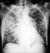
Chest radiograph showing extensive and coalescent larger acinar opacities in bronchopneumonia
Figure 7 (A and B)
Figure 7 (A and B)
A 40-year-old patient with acute onset of fever with dyspnea, not resolving with antibiotics. (A) Interstitial pneumonia in the form of well-defined consolidation, ill-defined scattered acinar infiltrates with air bronchograms (asterisk), GGO, and diffuse micronodular pattern. (B) Adenopathy (open arrow) seen on mediastinal window. There were microabscesses in the liver and spleen and ascites. The serology was positive for scrub typhus. Significant reduction in the lung lesions was seen after tetracycline therapy
Figure 8
Figure 8
A 40-year-old patient with fever, dry cough, headache, and dyspnea. Bilateral consolidation and GGO with subpleural and peribronchovascular predominance is seen. Throat swab was positive for H1N1
Figure 9
Figure 9
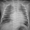
Chest radiograph in an infant with fever and respiratory distress shows features of bronchiolitis as hyperinflated lung fields and fine linear opacities due to atelectasis
Figure 10
Figure 10
A 12-year-old child with septicemia. Multiple well-defined nodules, more in subpleural region and most of them show a “feeding vessel sign” (arrow), suggesting septic emboli. Cavitation is seen in many nodules
Figure 11
Figure 11
Supine and prone axial CT scans showing “air crescent” sign of a dependent fungal ball in old lung cavity
Figure 12
Figure 12
Coronal reconstructed CT image showing fluid filled branching bronchoceles or “gloved finger sign” in ABPA
Figure 13
Figure 13
Serpentine old calcified guinea worm in muscles of the chest wall in a 60-year-old female overlying the right lung shadow
Figure 14
Figure 14
Bilateral dependent consolidation with air bronchogram. There is an anteroposterior gradient from normal lung to GGO to consolidation suggesting ARDS in the appropriate clinical setting
Figure 15
Figure 15
Wedge-shaped subpleural GGO in both lung apices. CT pulmonary angiogram in the revealed right pulmonary artery thrombosis
Figure 16
Figure 16
“CT angiogram sign.” Lobar consolidation with homogenous low attenuation and visibility of pulmonary vasculature through it
Figure 17
Figure 17
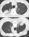
A 50-year-old male patient with dry cough for 6 months. Patchy wedge-shaped GGO which tend to be subpleural in location. The S. IgE levels in this patient were markedly raised upto 1818 kU/L, suggestive of chronic eosinophilic pneumonia
Figure 18 (A and B)
Figure 18 (A and B)
A 40-year-old male patient with cough and hemoptysis. (A, B) Combination of peripheral wedge-shaped GGO, consolidation, and cavity in left upper lobe (arrow), indicating a pulmonary vasculitis. ANCA was positive suggestive of granulomatous polyangitis
Figure 19 (A and B)
Figure 19 (A and B)
A 5-year-old male child with recurrent right lower lobe pneumonia and episode of massive hemoptysis. (A) Mass-like consolidation with cavitation in right lower lobe, with caudal extension into the retroperitoneum. (B) CT angiography shows an arterial feeder from the celiac artery (arrow). Radionuclide examination was positive for ectopic gastric mucosa indicating an infected transdiaphragmatic duplication cyst
Figure 20 (A and B)
Figure 20 (A and B)
(A, B) Imaging algorithm for approach to differential diagnosis of infectious pneumonia in immunocompetent host

Similar articles

References

    1. Ghanshani R, Gupta R, Gupta B, Kalra S, Khedar R, Sood S. Epidemiological study of prevalence, determinants, and outcomes of infections in medical ICU at a tertiary care hospital in India. Lung India. 2015;32:441. - PMC - PubMed
    1. Mandell L, Wunderink R, Anzueto A, Bartlett J, Campbell G, Dean N, et al. Infectious Diseases Society of America/American Thoracic Society Consensus Guidelines on the Management of Community-Acquired Pneumonia in Adults. Clin Infect Dis. 2007;44(Suppl 2):S27–72. - PMC - PubMed
    1. Gupta D, Agarwal R, Aggarwal A, Singh N, Mishra N, Khilnani G, et al. Guidelines for diagnosis and management of community-and hospital-acquired pneumonia in adults: Joint ICS/NCCP(I) recommendations. Lung India. 2012;29:27. - PMC - PubMed
    1. Guidelines for the Management of Adults with Hospital-acquired, Ventilator-associated, and Healthcare-associated Pneumonia. Am J Respir Crit Care Med. 2005;171:388–416. - PubMed
    1. Dandagi G. Nosocomial pneumonia in critically ill patients. Lung India. 2010;27:149. - PMC - PubMed