

Utility of supplemental screening with breast ultrasound in asymptomatic women with dense breast tissue who are not at high risk for breast cancer
- PMID: 28515586
- PMCID: PMC5385776
- DOI: 10.4103/0971-3026.202962
Utility of supplemental screening with breast ultrasound in asymptomatic women with dense breast tissue who are not at high risk for breast cancer
Abstract
Objective: To assess the results of an initial round of supplemental screening with hand-held bilateral breast ultrasound following a negative screening mammogram in asymptomatic women with dense breast tissue who are not at high risk for breast cancer.
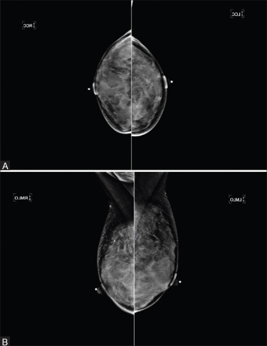
Materials and methods: A retrospective, Health Insurance Portability and Accountability Act compliant, Institutional Research Board approved study was performed at a single academic tertiary breast center. Informed consent was waived. A systematic review of the breast imaging center database was conducted to identify and retrieve data for all asymptomatic women, who were found to have heterogeneously dense or extremely dense breast tissue on screening bilateral mammograms performed from July 1, 2010 through June 30, 2012 and who received a mammographic final assessment American College of Radiology's (ACR) Breast Imaging Reporting and Data System (BI-RADS) category 1 or BI-RADS category 2. Hand-held screening ultrasound was performed initially by a technologist followed by a radiologist. Chi-square and t-test were used and statistical significance was considered at P < 0.05.
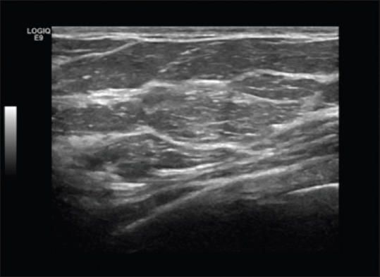
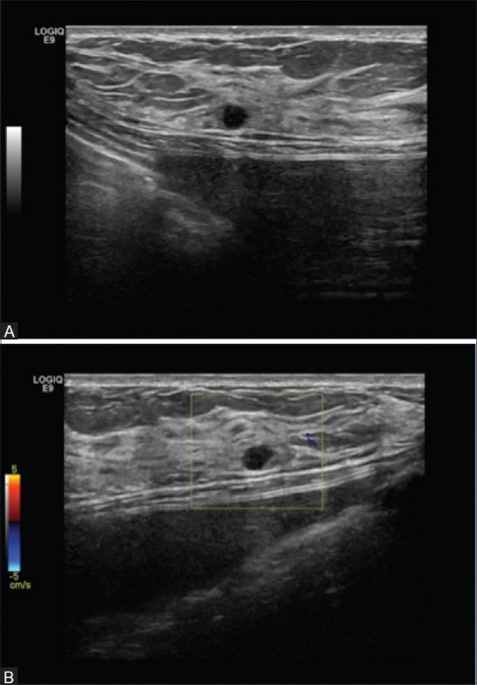
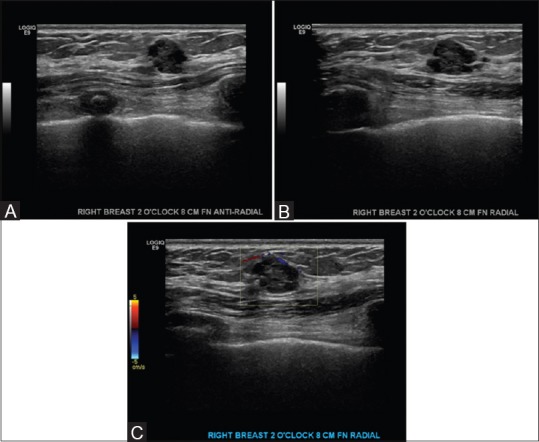
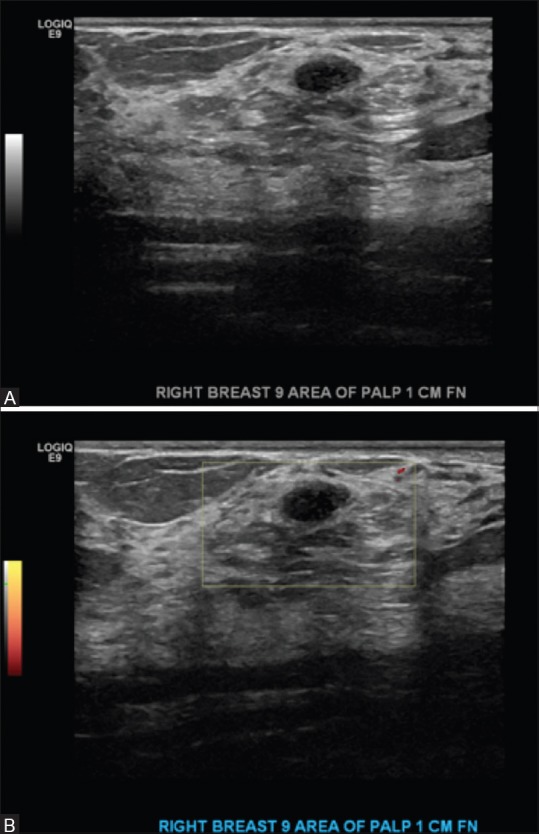
Results: A total of 1210 women were identified. Of these, 394 underwent the offered supplemental screening ultrasound. BI-RADS category 1 or 2 was assigned to 323 women (81.9%). BI-RADS category 3 was assigned to 50 women (12.9%). A total of 26 biopsies/aspirations were recommended and performed in 26 women (6.6%). The most common finding for which biopsy was recommended was a solid mass (88.5%) with an average size of 0.9 cm (0.5-1.7 cm). Most frequent pathology result was fibroadenoma (60.8%). No carcinoma was found.
Conclusion: Our data support the reported occurrence of a relatively high number of false positives at supplemental screening with breast ultrasound following a negative screening mammogram in asymptomatic women with dense breast tissue, who are not at a high risk of developing breast cancer, and suggests that caution is necessary in establishing wide implementation of this type of supplemental screening for all women with dense breast tissue without considering other risk factors for breast cancer.
Keywords: Breast ultrasound; dense breasts; screening.
Conflict of interest statement
There are no conflicts of interest.
Figures


References
-
- Breast Imaging Reporting and Data System. 4th ed. Reston: American College of Radiology; 2003.
-
- Carney PA, Miglioretti DL, Yankaskas BC, Kerlikowske K, Rosenberg R, Rutter CM, et al. Individual and combined effects of age, breast density, and hormone replacement therapy use on the accuracy of screening mammography. Ann Intern Med. 2003;138:168–75. - PubMed
-
- Boyd NF, Guo H, Martin LJ, Sun L, Stone J, Fishell E, et al. Mammographic density and the risk and detection of breast cancer. N Engl J Med. 2007;356:227–36. - PubMed
-
- Pisano ED, Gatsonis C, Hendrick E, Yaffe M, Baum JK, Acharyya S, et al. Digital Mammographic Imaging Screening Trial (DMIST) Investigators Group. Diagnostic performance of digital versus film mammography for breast-cancer screening. N Engl J Med. 2005;353:1773–83. - PubMed
LinkOut - more resources
Full Text Sources
Other Literature Sources