

Insulin Resistance as a Link between Amyloid-Beta and Tau Pathologies in Alzheimer's Disease
- PMID: 28515688
- PMCID: PMC5413582
- DOI: 10.3389/fnagi.2017.00118
Insulin Resistance as a Link between Amyloid-Beta and Tau Pathologies in Alzheimer's Disease
Abstract
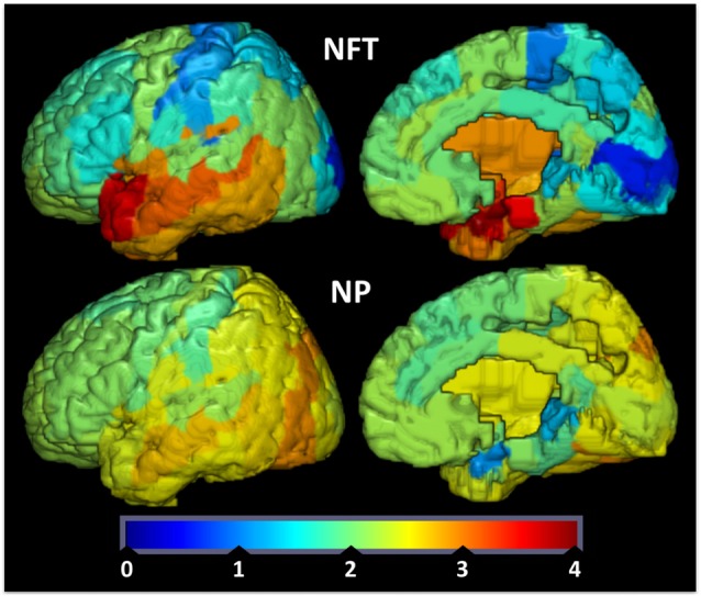
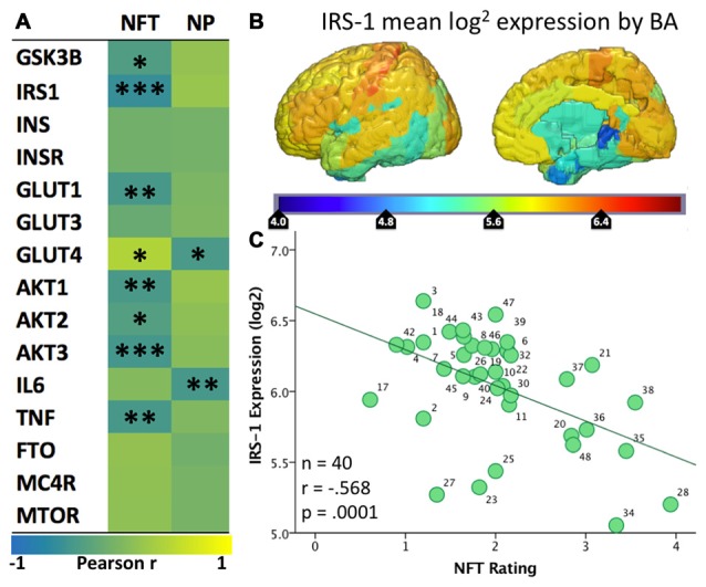
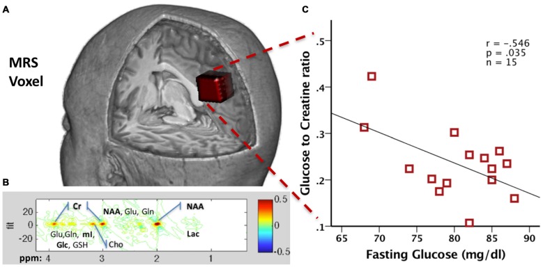
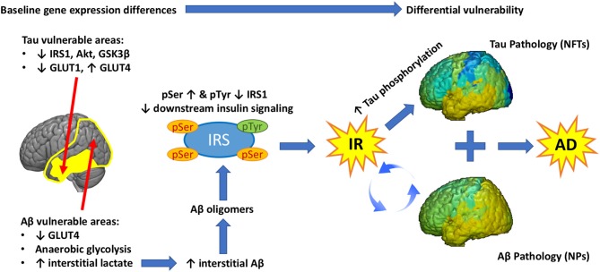
Current hypotheses and theories regarding the pathogenesis of Alzheimer's disease (AD) heavily implicate brain insulin resistance (IR) as a key factor. Despite the many well-validated metrics for systemic IR, the absence of biomarkers for brain-specific IR represents a translational gap that has hindered its study in living humans. In our lab, we have been working to develop biomarkers that reflect the common mechanisms of brain IR and AD that may be used to follow their engagement by experimental treatments. We present two promising biomarkers for brain IR in AD: insulin cascade mediators probed in extracellular vesicles (EVs) enriched for neuronal origin, and two-dimensional magnetic resonance spectroscopy (MRS) measures of brain glucose. As further evidence for a fundamental link between brain IR and AD, we provide a novel analysis demonstrating the close spatial correlation between brain expression of genes implicated in IR (using Allen Human Brain Atlas data) and tau and beta-amyloid pathologies. We proceed to propose the bold hypotheses that baseline differences in the metabolic reliance on glycolysis, and the expression of glucose transporters (GLUT) and insulin signaling genes determine the vulnerability of different brain regions to Tau and/or Amyloid beta (Aβ) pathology, and that IR is a critical link between these two pathologies that define AD. Lastly, we provide an overview of ongoing clinical trials that target IR as an angle to treat AD, and suggest how biomarkers may be used to evaluate treatment efficacy and target engagement.
Keywords: Alzheimer’s disease; IRS-1; exosomes; insulin resistance; magnetic resonance spectroscopy.
Figures
Similar articles
-
Decoding Alzheimer's disease from perturbed cerebral glucose metabolism: implications for diagnostic and therapeutic strategies.Prog Neurobiol. 2013 Sep;108:21-43. doi: 10.1016/j.pneurobio.2013.06.004. Epub 2013 Jul 11. Prog Neurobiol. 2013. PMID: 23850509 Review.
-
Insulin Resistance is Associated with Increased Levels of Cerebrospinal Fluid Biomarkers of Alzheimer's Disease and Reduced Memory Function in At-Risk Healthy Middle-Aged Adults.J Alzheimers Dis. 2016 Apr 12;52(4):1373-83. doi: 10.3233/JAD-160110. J Alzheimers Dis. 2016. PMID: 27079723 Free PMC article.
-
Amyloid-β and islet amyloid pathologies link Alzheimer's disease and type 2 diabetes in a transgenic model.FASEB J. 2017 Dec;31(12):5409-5418. doi: 10.1096/fj.201700431R. Epub 2017 Aug 14. FASEB J. 2017. PMID: 28808140
-
Insulin Resistance is Associated with Higher Cerebrospinal Fluid Tau Levels in Asymptomatic APOEɛ4 Carriers.J Alzheimers Dis. 2015;46(2):525-33. doi: 10.3233/JAD-150072. J Alzheimers Dis. 2015. PMID: 25812851 Free PMC article.
-
Insulin resistance in Alzheimer's disease.Transl Res. 2017 May;183:26-40. doi: 10.1016/j.trsl.2016.12.005. Epub 2016 Dec 13. Transl Res. 2017. PMID: 28034760 Free PMC article. Review.
Cited by
-
Blueberry Supplementation in Midlife for Dementia Risk Reduction.Nutrients. 2022 Apr 13;14(8):1619. doi: 10.3390/nu14081619. Nutrients. 2022. PMID: 35458181 Free PMC article. Clinical Trial.
-
Blood-brain barrier permeability and physical exercise.J Neuroinflammation. 2019 Jan 24;16(1):15. doi: 10.1186/s12974-019-1403-x. J Neuroinflammation. 2019. PMID: 30678702 Free PMC article. Review.
-
Anti-Amyloidogenic Effects of Metasequoia glyptostroboides Fruits and Its Active Constituents.Molecules. 2023 Jan 19;28(3):1017. doi: 10.3390/molecules28031017. Molecules. 2023. PMID: 36770688 Free PMC article.
-
Structural and functional imaging of brains.Sci China Chem. 2023;66(2):324-366. doi: 10.1007/s11426-022-1408-5. Epub 2022 Dec 9. Sci China Chem. 2023. PMID: 36536633 Free PMC article. Review.
-
Neddylation-dependent protein degradation is a nexus between synaptic insulin resistance, neuroinflammation and Alzheimer's disease.Transl Neurodegener. 2022 Jan 6;11(1):2. doi: 10.1186/s40035-021-00277-8. Transl Neurodegener. 2022. PMID: 34986876 Free PMC article.
References
-
- Apelt J., Mehlhorn G., Schliebs R. (1999). Insulin-sensitive GLUT4 glucose transporters are colocalized with GLUT3-expressing cells and demonstrate a chemically distinct neuron-specific localization in rat brain. J. Neurosci. Res. 57, 693–705. 10.1002/(sici)1097-4547(19990901)57:5<693::aid-jnr11>3.0.co;2-x - DOI - PubMed
-
- Arnold S. E., Hyman B. T., Flory J., Damasio A. R., Van Hoesen G. W. (1991). The topographical and neuroanatomical distribution of neurofibrillary tangles and neuritic plaques in the cerebral cortex of patients with Alzheimer’s disease. Cereb. Cortex 1, 103–116. 10.1093/cercor/1.1.103 - DOI - PubMed
LinkOut - more resources
Full Text Sources
Other Literature Sources
Research Materials
Miscellaneous