Review
. 2017 May 3:9:129.
doi: 10.3389/fnagi.2017.00129. eCollection 2017.

Brain Renin-Angiotensin System and Microglial Polarization: Implications for Aging and Neurodegeneration

Affiliations
Review

Brain Renin-Angiotensin System and Microglial Polarization: Implications for Aging and Neurodegeneration

Jose L Labandeira-Garcia et al. Front Aging Neurosci. .

Abstract

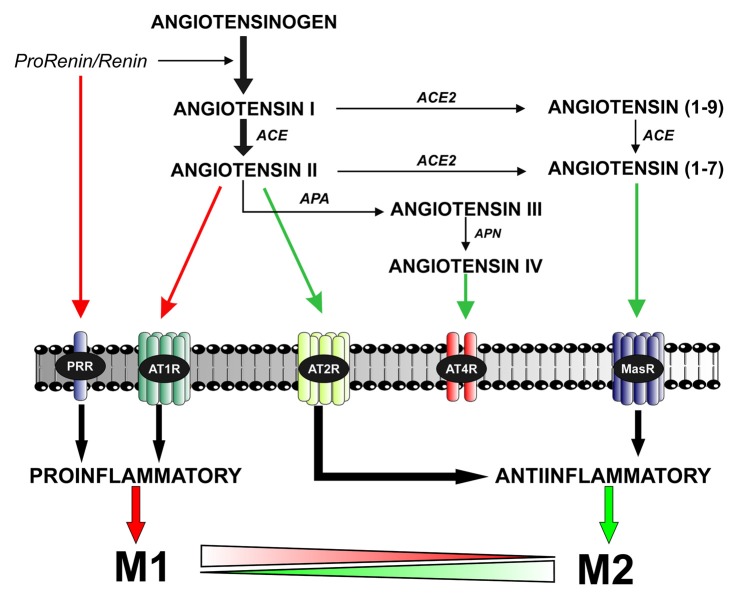
Microglia can transform into proinflammatory/classically activated (M1) or anti-inflammatory/alternatively activated (M2) phenotypes following environmental signals related to physiological conditions or brain lesions. An adequate transition from the M1 (proinflammatory) to M2 (immunoregulatory) phenotype is necessary to counteract brain damage. Several factors involved in microglial polarization have already been identified. However, the effects of the brain renin-angiotensin system (RAS) on microglial polarization are less known. It is well known that there is a "classical" circulating RAS; however, a second RAS (local or tissue RAS) has been observed in many tissues, including brain. The locally formed angiotensin is involved in local pathological changes of these tissues and modulates immune cells, which are equipped with all the components of the RAS. There are also recent data showing that brain RAS plays a major role in microglial polarization. Level of microglial NADPH-oxidase (Nox) activation is a major regulator of the shift between M1/proinflammatory and M2/immunoregulatory microglial phenotypes so that Nox activation promotes the proinflammatory and inhibits the immunoregulatory phenotype. Angiotensin II (Ang II), via its type 1 receptor (AT1), is a major activator of the NADPH-oxidase complex, leading to pro-oxidative and pro-inflammatory effects. However, these effects are counteracted by a RAS opposite arm constituted by Angiotensin II/AT2 receptor signaling and Angiotensin 1-7/Mas receptor (MasR) signaling. In addition, activation of prorenin-renin receptors may contribute to activation of the proinflammatory phenotype. Aged brains showed upregulation of AT1 and downregulation of AT2 receptor expression, which may contribute to a pro-oxidative pro-inflammatory state and the increase in neuron vulnerability. Several recent studies have shown interactions between the brain RAS and different factors involved in microglial polarization, such as estrogens, Rho kinase (ROCK), insulin-like growth factor-1 (IGF-1), tumor necrosis factor α (TNF)-α, iron, peroxisome proliferator-activated receptor gamma, and toll-like receptors (TLRs). Metabolic reprogramming has recently been involved in the regulation of the neuroinflammatory response. Interestingly, we have recently observed a mitochondrial RAS, which is altered in aged brains. In conclusion, dysregulation of brain RAS plays a major role in aging-related changes and neurodegeneration by exacerbation of oxidative stress (OS) and neuroinflammation, which may be attenuated by pharmacological manipulation of RAS components.

Keywords: NADPH-oxidase; Nox; Parkinson; angiotensin; microglia; neuroinflammation; neuroprotection; oxidative stress.


Figures

Figure 1
Figure 1
The brain renin–angiotensin system (RAS). Angiotensin II is formed by the sequential action of two enzymes prorenin-renin (that forms angiotensin I) and ACE on the precursor protein angiotensinogen. Renin and its precursor prorenin also act on specific PRR receptors. In addition to angiotensin II, several angiotensin peptides such as Angiotensin (1–7), angiotensin III and angiotensin IV are formed from angiotensin I and II by the action of ACE2, APA and APN. Angiotensin II, via AT1 receptors, induces pro-inflammatory effects and M1 phenotype. However, these effects are counteracted by a RAS opposite arm constituted by angiotensin II/AT2 receptor signaling and Angiotensin 1–7/MasR signaling. In addition, activation of prorenin-renin receptors may contribute to activation of the proinflammatory phenotype. Abbreviations: ACE, angiotensin converting enzyme; ACE2, angiotensin converting enzyme 2; APA, aminopeptidase A; APN, aminopeptidase N; AT1R, angiotensin type 1 receptor; AT2R, angiotensin type 2 receptor; AT4R, angiotensin type 4 receptor; MasR, Mas receptor; PRR, prorenin/renin receptor.
Figure 2
Figure 2
Intense microglial activation in a degenerating rat substantia nigra (A), iba-1 positive red immunofluorescence. A high density of activated microglial cells is observed in the nigral region surrounding the dopaminergic cells. The degeneration of dopaminergic neurons (green; tyrosine hydroxylase, TH immunolabeled cells) was triggered by over expression of alpha-synuclein (not shown) after unilateral injection of adeno-associated viral vectors encoding alpha-synuclein. The contralateral non-lesioned substantia nigra of the same tissue section (B) shows scattered microglial cells in an apparent classical “non-activated” state. The areas squared in (A,B) are magnified in (C–E), and a close interaction between a dopaminergic neuron and a microglial cell (asterisks) is shown in (E). Scale bar: 100 μm (A,B), 20 μm (C,D) and 10 μm (E).
Figure 3
Figure 3
Dysregulation of brain RAS with aging and other pathogenic factors. In young and healthy brains the balance between the RAS pro-oxidative/proinflammatory and protective arms are tight regulated for physiological functions. Aging is associated with overactivation of the Ras pro-oxidative/proinflammatory arm and lack of compensatory upregulation of the protective arm, which leads to a pro-oxidative pro-inflammatory state and increased vulnerability to neurodegeneration. Other pathogenic factors involved in triggering neurodegeneration may also increase the activity of the RAS pro-inflammatory axis. Abbreviations: Ang, angiotensin; AT1, angiotensin type 1 receptor; AT2, angiotensin type 2 receptor; ND, neurodegenerative; Nox, NADPH-oxidase; OS, oxidative stress; PRR, prorenin/renin receptor.
Figure 4
Figure 4
Role of different angiotensin receptors in regulation of microglial polarization, and interactions with other microglial polarization regulators. Angiotensin II, via AT1 receptor and Nox activation, and prorenin/renin, via PRR, induce pro-inflammatory effects and M1 phenotype, which is associated to increased levels of ROS, NO, TNF-α and IL-1β. These effects are counteracted by a RAS opposite arm constituted by angiotensin II/AT2 receptor signaling and Angiotensin 1–7/MasR signaling, which promote anti-inflammatory effects and M2 phenotype, which is also associated to TGF-β, IL-4, IL-10, and Arg-1 overexpression. In addition, there are interactions between RAS and different compounds involved in neuroinflammation and microglial polarization, such as estrogens, ROCK, IGF1, TNF-α, iron, PPARγ, and toll-like receptors. Abbreviations: Ang, angiotensin; ARG-1, arginase 1; AT1, angiotensin type 1 receptor; AT2, angiotensin type 2 receptor; IGF1, insulin-like growth factor-1; IL, interleukin; MasR, Mas receptor; NO, nitric oxide; Nox, NADPH-oxidase; O2, superoxide; PPARγ, peroxisome proliferator-activated receptor gamma; PRR, prorenin/renin receptor; ROCK, Rho kinase; ROS, reactive oxygen species; TGF-β, transforming growth factor β; TNF-α tumor necrosis factor α. Scale bar: 7.5 μm.

References

    1. Alberici L. C., Oliveira H. C., Paim B. A., Mantello C. C., Augusto A. C., Zecchin K. G., et al. . (2009). Mitochondrial ATP-sensitive K+ channels as redox signals to liver mitochondria in response to hypertriglyceridemia. Free Radic. Biol. Med. 47, 1432–1439. 10.1016/j.freeradbiomed.2009.08.013 - DOI - PubMed
    1. Albiston A. L., McDowall S. G., Matsacos D., Sim P., Clune E., Mustafa T., et al. . (2001). Evidence that the angiotensin IV (AT4) receptor is the enzyme insulin-regulated aminopeptidase. J. Biol. Chem. 276, 48623–48626. 10.1074/jbc.c100512200 - DOI - PubMed
    1. Alliot F., Godin I., Pessac B. (1999). Microglia derive from progenitors, originating from the yolk sac, and which proliferate in the brain. Dev. Brain Res. 117, 145–152. 10.1016/s0165-3806(99)00113-3 - DOI - PubMed
    1. Babior B. M. (2004). NADPH oxidase. Curr. Opin. Immunol. 16, 42–47. 10.1016/j.coi.2003.12.001 - DOI - PubMed
    1. Bacani C., Frishman W. H. (2006). Chymase: a new pharmacologic target in cardiovascular disease. Cardiol. Rev. 14, 187–193. 10.1097/01.crd.0000195220.62533.c5 - DOI - PubMed