. 2017 May 16;6(3):4.
doi: 10.1167/tvst.6.3.4. eCollection 2017 May.

Transplantation of Human Embryonic Stem Cell-Derived Retinal Cells into the Subretinal Space of a Non-Human Primate

Affiliations

Transplantation of Human Embryonic Stem Cell-Derived Retinal Cells into the Subretinal Space of a Non-Human Primate

Jennifer R Chao et al. Transl Vis Sci Technol. .

Abstract

Purpose: Previous studies have demonstrated the ability of retinal cells derived from human embryonic stem cells (hESCs) to survive, integrate into the host retina, and mediate light responses in murine mouse models. Our aim is to determine whether these cells can also survive and integrate into the retina of a nonhuman primate, Saimiri sciureus, following transplantation into the subretinal space.

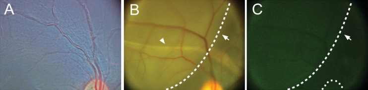
Methods: hESCs were differentiated toward retinal neuronal fates using our previously published technique and cultured for 60 to 70 days. Differentiated cells were further treated with 20 μM N-[N-(3,5-Difluorophenacetyl)-L-alanyl]-S-phenylglycine t-butyl ester (DAPT) for a period of 5 days immediately prior to subretinal transplantation. Differentiated cells were labeled with a lentivirus expressing GFP. One million cells (10,000 cells/μL) were injected into the submacular space into a squirrel monkey eye, using an ab externo technique.

Results: RetCam imaging demonstrated the presence and survival of human donor cells 3 months after transplantation in the S. sciureus eye. Injected cells consolidated in the temporal macula. GFP+ axonal projections were observed to emanate from the central consolidation of cells at 1 month, with some projecting into the optic nerve by 3 months after transplantation.

Conclusions: Human ES cell-derived retinal neurons injected into the submacular space of a squirrel monkey survive at least 3 months postinjection without immunosuppression. Some donor cells appeared to integrate into the host inner retina, and numerous donor axonal projections were noted throughout, with some projecting into the optic nerve.

Translational relevance: These data illustrate the feasibility of hESC-derived retinal cell replacement in the nonhuman primate eye.

Keywords: AMD; glaucoma; primate; retina; stem cell transplantation.


Figures

Figure 1
Figure 1
DAPT induces retinal progenitor differentiation and inhibits proliferation. hESCs were differentiated toward retinal neuronal fates. (A) Brightfield and immunostaining of control and DAPT-treated cells after 5 days. A decrease in the number of rosettes, LHX2 and PAX6 expression, along with decreased PH3 staining of mitotic cells was observed in DAPT-treated cells. (B) DAPT treatment resulted in increased TUJ1- and HuC/D-expressing retinal cells. (C) Immunostaining reveals retinal neurons expressing photoreceptor markers, recoverin, and OTX2 (arrows). (D) RT-PCR analysis demonstrated decreased PAX6 and increased BRN3B and RCVRN expression in DAPT-treated cells compared to DMSO controls. Mean ± SEM, *P < 0.05, **P < 0.01, ***P < 0.005.
Figure 2
Figure 2
Ab externo delivery of hESC-derived retinal cells into the subretinal space of the Saimiri sciureus eye. (A) A radial sclerotomy was created and the incision was spread with a microretractor. (B) Dissection of choroid fibers and a choroidotomy was created using a wire-tipped cannula (arrowhead). A small amount of Healon was infused through the choroidotomy to create a subretinal bleb. (C) The subretinal catheter with a lighted fiberoptic tip (arrowhead) was inserted at a low angle into the subretinal space. (D) The catheter was advanced along the subretinal space into the superior macula, and the lighted (red) catheter tip could be visualized through a contact lens (arrowhead). Optic nerve (outlined by dotted line, D). The subretinal injection was performed under direct visualization.
Figure 3
Figure 3
hESC-derived retinal cells transplanted into the superior macula. One million cells in a volume of 100 μL (10,000 cells/μL) were injected into the subretinal space of the superior macula. (A) Preinjection image of the superior macula and optic nerve. Donor cells were transduced with a lentivirus expressing EF1α-GFP. (B) Immediately postinjection, a localized subretinal detachment at the subretinal cannula infusion extending from the superior arcade into the central macula (dotted line, arrow). No retinal holes or tears were identified. The subretinal catheter track of RPE hypopigmentation could be appreciated (arrowhead). (C) GFP+-injected cells could be visualized in the subretinal space with RetCam imaging (dotted line, arrow). Optic nerve is delineated (dotted line).
Figure 4
Figure 4
Transplanted hESC-derived retinal cells survive and extend axonal projections. (A) At 4 weeks, the injected hESC-derived retinal cells were observed to have consolidated in the posterior pole temporal to the fovea, corresponding to the most inferior and posterior extent of the original subretinal injection. (B) At 8 weeks postinjection, axonal projections emanating from the subretinal cell mass were observed, with some extending toward the optic nerve (arrow). (C) Additional projections were noted 12 weeks postinjection (arrow), with some projections appearing to change direction to travel along arcuate nerve fiber bundles with the host retinal NFL. Despite the absence of immunosuppression, there was no evidence of anterior or posterior segment inflammation at any time posttransplantation.
Figure 5
Figure 5
Transplanted cells migrate into inner retinal cell layers and extend axonal projections into the optic nerve. (A) At 12 weeks postinjection, few GFP+ cells were observed in the inner nuclear layer (arrowhead) and GCL (arrow). (B) Immunostaining of the subretinal mass of nonintegrated cells revealed few Ki-67+ cells (arrow). (C) Axonal projections emanating from the transplanted cells extended along the inner plexiform layer, GCL, and NFL. GFP+ projections were noted entering at the optic nerve head (D, inset and D, arrow), and extending into the retrolaminar optic nerve (D-D”). ON, optic nerve.
Figure 6
Figure 6
GFP+ cells and axonal extensions co-label with the human specific cell marker, STEM121. (A) Immunostaining at 12 weeks postinjection reveals GFP+ axons in the host NFL (arrow), as well as nonanatomical axonal projections through the inner plexiform layer and GCL (open arrowhead). GFP+ axonal extensions co-localize with STEM121 (human specific marker) immunostaining (A’ and A”, arrow), but some STEM121+ axonal extensions do not co-localize with the GFP signal (A’ and A”, closed arrowhead). (B-B’”) GFP+/STEM121+ axons are observed in the host NFL, which is strongly labeled with TUJ1 (arrow). (C-C’”) TUJ1 co-labeling of donor GFP+/STEM121+ axons (arrows) is evident in the outer plexiform layer where there are comparatively lower levels of TUJ1 expression in the host retina. (D-D”) A few GFP+ cells that co-label with STEM121 are located in the GCL and extend axonal projections into the NFL of the host retina. INL, inner nuclear layer; IPL, inner plexiform layer; ONL, outer nuclear layer; OPL, outer plexiform layer.

References

    1. Ambati J,, Fowler BJ. Mechanisms of age-related macular degeneration. Neuron. 2012; 75: 26–39. - PMC - PubMed
    1. Klein R,, Chou CF,, Klein BE,, Zhang X,, Meuer SM,, Saaddine JB. Prevalence of age-related macular degeneration in the US population. Arch Ophthalmol. 2011; 129: 75–80. - PubMed
    1. Banin E,, Obolensky A,, Idelson M,, et al. Retinal incorporation and differentiation of neural precursors derived from human embryonic stem cells. Stem Cells. 2006; 24: 246–257. - PubMed
    1. Gonzalez-Cordero A,, West EL,, Pearson RA,, et al. Photoreceptor precursors derived from three-dimensional embryonic stem cell cultures integrate and mature within adult degenerate retina. Nat Biotechnol. 2013; 31: 741–747. - PMC - PubMed
    1. Lamba DA,, Gust J,, Reh TA. Transplantation of human embryonic stem cell-derived photoreceptors restores some visual function in Crx-deficient mice. Cell Stem Cell. 2009; 4: 73–79. - PMC - PubMed