Review
. 2013 Apr 1;1(1):e24360.
doi: 10.4161/idp.24360. eCollection 2013 Jan-Dec.

The alphabet of intrinsic disorder: I. Act like a Pro: On the abundance and roles of proline residues in intrinsically disordered proteins

Affiliations
Review

The alphabet of intrinsic disorder: I. Act like a Pro: On the abundance and roles of proline residues in intrinsically disordered proteins

Francois-Xavier Theillet et al. Intrinsically Disord Proteins. .

Abstract

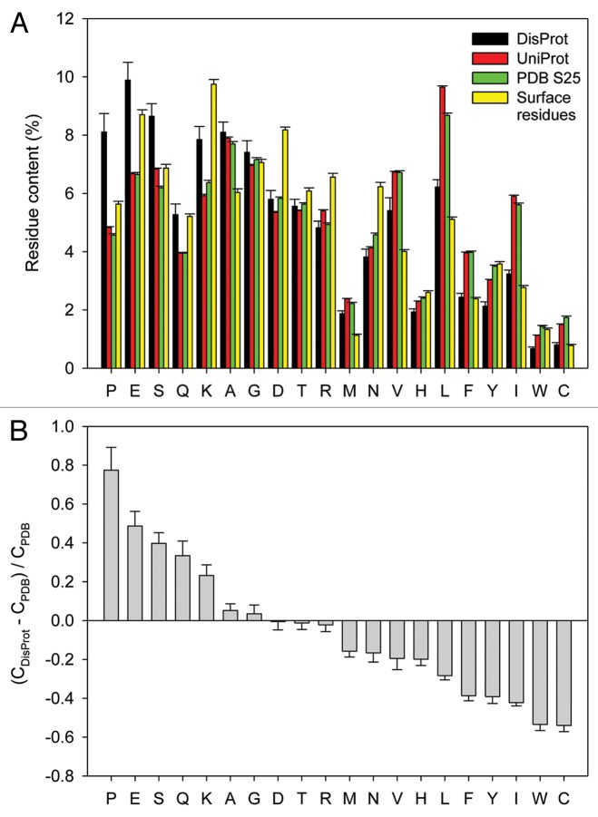
A significant fraction of every proteome is occupied by biologically active proteins that do not form unique three-dimensional structures. These intrinsically disordered proteins (IDPs) and IDP regions (IDPRs) have essential biological functions and are characterized by extensive structural plasticity. Such structural and functional behavior is encoded in the amino acid sequences of IDPs/IDPRs, which are enriched in disorder-promoting residues and depleted in order-promoting residues. In fact, amino acid residues can be arranged according to their disorder-promoting tendency to form an alphabet of intrinsic disorder that defines the structural complexity and diversity of IDPs/IDPRs. This review is the first in a series of publications dedicated to the roles that different amino acid residues play in defining the phenomenon of protein intrinsic disorder. We start with proline because data suggests that of the 20 common amino acid residues, this one is the most disorder-promoting.

Keywords: cis-trans isomerization; conformational restriction; intrinsically disordered protein; post-translational modification; protein solubility; protein surfaces.


Figures

None
Figure 1. Amino acid determinants defining structural and functional differences between the ordered and intrinsically disordered proteins. (A) Amino acid compositions of several data sets discussed in the text (DisProt, UniProt, PDB Select 25 and surface residues37). (B) Fractional difference in the amino acid composition (compositional profile) between the typical IDPs from the DisProt database and a set of completely ordered proteins calculated for each amino acid residue. The fractional difference was evaluated as (CDisProt-CPDB)/CPDB, where CDisProt is the content of a given amino acid in a DisProt databse, and CPDB is the corresponding content in the data set of fully ordered proteins. Positive bars correspond to residues found more abundantly in IDPs, whereas negative bars show residues, in which IDPs are depleted. Amino acid types were ranked according to their decreasing disorder-promoting potential.
None
Figure 2. Chemical structure of peptide fragments in trans (A) and (C) and cis conformation (B) and (D); (C) and (D) show a proline-containing fragment. The red arrows point out the steric hindrances between the Cα of the residue (−1) with the Hamide (A) or the Cα of the residue (0) (B) for the non-proline-containing peptides, and between the Cα of the residue (-1) with the Cδ (C) or the Cα of the proline (D). Ramachandran plots of non-proline, non-glycine, non-isoleucine, non-valine residues (E) and proline residues (F) result from the analysis of 1.5 million residues in 8,000 protein chains with resolution < 2 Å and backbone B-factors < 30.The contours separate the “outlier,” “allowed” and “favored” regions of the Ramachandran plots. The Ramachandran plots were adapted from commons.wikimedia.org/wiki/User:Dcrjsr. The β-strand (β), α-helix (α), α-L-helix (αL), poly-proline II (PPII) regions of the Ramachandran plots are indicated and we show a representation of a model poly-proline II helix.
None
Figure 3. A two-dimensional plot correlating proline and glycine content for a wide variety of elastomeric and amyloidogenic peptides. Elastomeric proteins are characterized by high GP content and are located in the upper-right part of this plot. Contrarily, amyloidogenic peptides are characterized by low PG content and therefore are located in the left bottom corner of the plot. The coexistence region (shaded in gray) contains P and G compositions consistent with both amyloidogenic and elastomeric properties. Elastomeric proteins, including the domains of elastin, major ampullate spindroin (MaSp) 2, flagelliform silk, the elastic domains of mussel byssus thread, and abductin, appear above a composition threshold (upper dashed line). Amyloidogenic sequences are primarily found below the PG-threshold, along with rigid lizard egg shells, tubulliform silk (TuSp1), a protective silk for spider eggs, and aciniform silk (AcSp), used for wrapping prey. The coexistence region contains amyloid-like peptides as well as the elastomeric adhesive produced by the frog Notaden bennetti, the PEVK domains of titin, wheat glutenin protein, and the strongest spider silks, namely MaSp1 and minor ampullate spindroin (MiSp). Figure reproduced from ref. Abbreviations: AcSp, aciniform silk; MaSp, major ampullate spindroin; MiSp, minor ampullate spindroin; TuSp1, tubulliform silk.

References

    1. Uversky VN. . Intrinsically disordered proteins from A to Z. Int J Biochem Cell Biol 2011; 43:1090 - 103; http://dx.doi.org/ 10.1016/j.biocel.2011.04.001; PMID: 21501695 - DOI - PubMed
    1. Tompa P. . Unstructural biology coming of age. Curr Opin Struct Biol 2011; 21:419 - 25; http://dx.doi.org/ 10.1016/j.sbi.2011.03.012; PMID: 21514142 - DOI - PubMed
    1. Romero P, Obradovic Z, Kissinger CR, Villafranca JE, Garner E, Guilliot S, et al. . Thousands of proteins likely to have long disordered regions. Pac Symp Biocomput 1998; •••:437 - 48; PMID: 9697202 - PubMed
    1. Dunker AK, Obradovic Z, Romero P, Garner EC, Brown CJ. . Intrinsic protein disorder in complete genomes. Genome Inform Ser Workshop Genome Inform 2000; 11:161 - 71; PMID: 11700597 - PubMed
    1. Uversky VN, Gillespie JR, Fink AL. . Why are “natively unfolded” proteins unstructured under physiologic conditions?. Proteins 2000; 41:415 - 27; http://dx.doi.org/ 10.1002/1097-0134(20001115)41:3<415::AID-PROT130>3.0.CO;2-7; PMID: 11025552 - DOI - PubMed