

A method to trap transient and weak interacting protein complexes for structural studies
- PMID: 28516014
- PMCID: PMC5424782
- DOI: 10.4161/idp.25464
A method to trap transient and weak interacting protein complexes for structural studies
Abstract
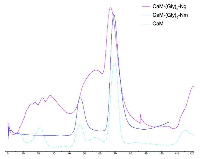
Several key biological events adopt a "hit-and-run" strategy in their transient interactions between binding partners. In some instances, the disordered nature of one of the binding partners severely hampers the success of co-crystallization, often leading to the crystallization of just one of the partners. Here, we discuss a method to trap weak and transient protein interactions for crystallization. This approach requires the structural details of at least one of the interacting partners and binding knowledge to dock the known minimum binding region (peptide) of the protein onto the other using an optimal-sized linker. Prior to crystallization, the purified linked construct should be verified for its intact folding and stability. Following structure determination, structure-guided functional studies are performed with independent, full-length unlinked proteins to validate the findings of the linked complex. We designed this approach and then validated its efficacy using a 24 amino acid minimum binding region of the intrinsically disordered, neuron-specific substrates, Neurogranin and Neuromodulin, joined via a Gly-linker to their interacting partner, Calmodulin. Moreover, the reported functional studies with independent full-length proteins confirmed the findings of the linked peptide complexes. Based on our studies, and in combination with the supporting literature, we suggest that optimized linkers can provide an environment to mimic the natural interactions between binding partners, and offer a useful strategy for structural studies to trap weak and transient interactions involved in several biological processes.
Keywords: Protein-protein interactions; co-crystallization; linked complex; transient binding.
Figures


References
-
- Ozbabacan SE, Engin HB, Gursoy A, Keskin O. . Transient protein-protein interactions. Protein Eng Des Sel 2011; 24:635 - 48; http://dx.doi.org/ 10.1093/protein/gzr025; PMID: 21676899 - DOI - PubMed
-
- Dyson HJ, Wright PE. . Coupling of folding and binding for unstructured proteins. Curr Opin Struct Biol 2002; 12:54 - 60; http://dx.doi.org/ 10.1016/S0959-440X(02)00289-0; PMID: 11839490 - DOI - PubMed
-
- Back JW, de Jong L, Muijsers AO, de Koster CG. . Chemical cross-linking and mass spectrometry for protein structural modeling. J Mol Biol 2003; 331:303 - 13; http://dx.doi.org/ 10.1016/S0022-2836(03)00721-6; PMID: 12888339 - DOI - PubMed
-
- Sinz A. . Chemical cross-linking and mass spectrometry to map three-dimensional protein structures and protein-protein interactions. Mass Spectrom Rev 2006; 25:663 - 82; http://dx.doi.org/ 10.1002/mas.20082; PMID: 16477643 - DOI - PubMed
LinkOut - more resources
Full Text Sources
Other Literature Sources