

Necrotizing Pancreatitis: Current Management and Therapies
- PMID: 28516758
- PMCID: PMC5565044
- DOI: 10.5946/ce.2016.152
Necrotizing Pancreatitis: Current Management and Therapies
Abstract
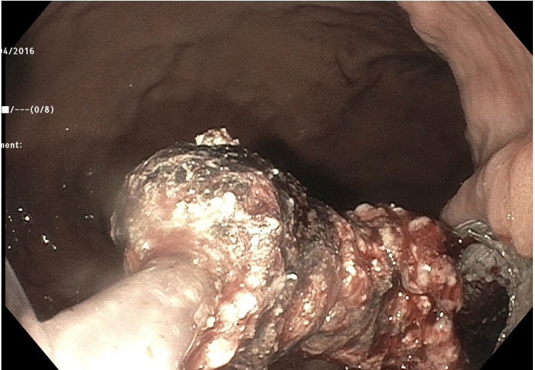
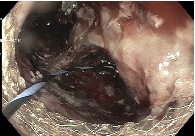
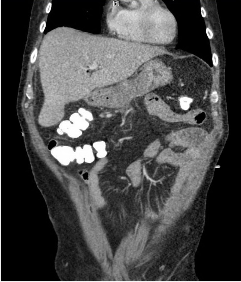
Acute necrotizing pancreatitis accounts for 10% of acute pancreatitis (AP) cases and is associated with a higher mortality and morbidity. Necrosis within the first 4 weeks of disease onset is defined as an acute necrotic collection (ANC), while walled off pancreatic necrosis (WOPN) develops after 4 weeks of disease onset. An infected or symptomatic WOPN requires drainage. The management of pancreatic necrosis has shifted away from open necrosectomy, as it is associated with a high morbidity, to less invasive techniques. In this review, we summarize the current management and therapies for acute necrotizing pancreatitis.
Keywords: Necrosis; Pancreatitis; Pseudocyst; Walled-off pancreatic necrosis.
Conflict of interest statement
Figures
References
-
- Banks PA, Bollen TL, Dervenis C, et al. Classification of acute pancreatitis--2012: revision of the Atlanta classification and definitions by international consensus. Gut. 2013;62:102–111. - PubMed
-
- Mofidi R, Duff MD, Wigmore SJ, Madhavan KK, Garden OJ, Parks RW. Association between early systemic inflammatory response, severity of multiorgan dysfunction and death in acute pancreatitis. Br J Surg. 2006;93:738–744. - PubMed
-
- Adler DG, Baron TH, Davila RE, et al. ASGE guideline: the role of ERCP in diseases of the biliary tract and the pancreas. Gastrointest Endosc. 2005;62:1–8. - PubMed
-
- Dervenis C, Johnson CD, Bassi C, et al. Diagnosis, objective assessment of severity, and management of acute pancreatitis. Santorini consensus conference. Int J Pancreatol. 1999;25:195–210. - PubMed
Publication types
LinkOut - more resources
Full Text Sources
Other Literature Sources
