. 2017 Jun 13;56(23):2938-2949.
doi: 10.1021/acs.biochem.6b01182. Epub 2017 May 25.

An Autoinhibitory Role for the Pleckstrin Homology Domain of Interleukin-2-Inducible Tyrosine Kinase and Its Interplay with Canonical Phospholipid Recognition

Affiliations

An Autoinhibitory Role for the Pleckstrin Homology Domain of Interleukin-2-Inducible Tyrosine Kinase and Its Interplay with Canonical Phospholipid Recognition

Sujan Devkota et al. Biochemistry. .

Abstract

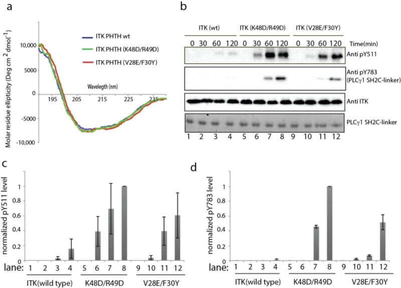
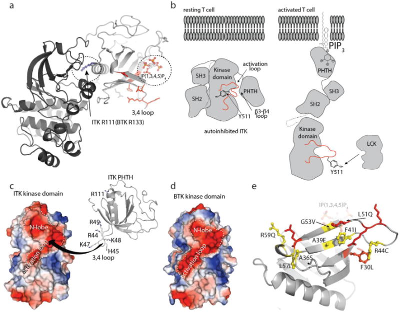
Pleckstrin homology (PH) domains are well-known as phospholipid binding modules, yet evidence that PH domain function extends beyond lipid recognition is mounting. In this work, we characterize a protein binding function for the PH domain of interleukin-2-inducible tyrosine kinase (ITK), an immune cell specific signaling protein that belongs to the TEC family of nonreceptor tyrosine kinases. Its N-terminal PH domain is a well-characterized lipid binding module that localizes ITK to the membrane via phosphatidylinositol 3,4,5-trisphosphate (PIP3) binding. Using a combination of nuclear magnetic resonance spectroscopy and mutagenesis, we have mapped an autoregulatory protein interaction site on the ITK PH domain that makes direct contact with the catalytic kinase domain of ITK, inhibiting the phospho-transfer reaction. Moreover, we have elucidated an important interplay between lipid binding by the ITK PH domain and the stability of the autoinhibitory complex formed by full length ITK. The ITK activation loop in the kinase domain becomes accessible to phosphorylation to the exogenous kinase LCK upon binding of the ITK PH domain to PIP3. By clarifying the allosteric role of the ITK PH domain in controlling ITK function, we have expanded the functional repertoire of the PH domain generally and opened the door to alternative strategies to target this specific kinase in the context of immune cell signaling.


Figures

Figure 1
Figure 1. The ITK PHTH domain interacts with the ITK kinase domain in trans
(a) Domain constructs of ITK used in this study. Full length ITK is shown above the His6-GB1-tagged PHTH domain fragment of ITK (residues 1-154) and the His6-tagged Kinase domain fragment (residues 356-619). (b) Coomassie gel showing ITK kinase domain (4 μM) associates with ITK PHTH domain (2 and 4 μM in lanes 7 and 8, respectively) immobilized on IgG sepharose beads. Lanes 1 shows the ITK kinase domain alone does not associate with IgG sepharose, lanes 2, 3 and 4 are loading controls. Lane 5 shows the ITK kinase domain and asterisks show the IgG heavy and light chains. Lane 6 shows that the ITK kinase domain does not associate with IgG sepharose bound His6-GB1 that lacks ITK PHTH. (c) Domain constructs of BTK used in this study. Full length BTK is shown above the His6-GB1-tagged PHTHSH3 (residues 1-270), the His6-GB1-tagged PHTH (residues 1-176) and the His6-tagged Kinase domain fragments of BTK (residues 396-659 (Kinase domain) and residues 382-659 (Linker-Kinase domain)). (d) Coomassie gel showing that neither the BTK kinase domain (1 μM) or the BTK linker-kinase domain (1 μM) associates in this assay with either the BTK PHTH domain (2 μM) or the larger BTK PHTHSH3 construct (2 μM) immobilized on IgG sepharose beads (lanes 6,7,9 and 10). Protein alone loading controls are shown on the left side of the gel. As in (b) asterisks indicate IgG heavy and light chains.
Figure 2
Figure 2. NMR mapping of the ITK PHTH regulatory interface
(a) [15N,1H]-HSQC spectrum of uniformly 15N-labeled ITK PHTH domain. (b,c) Addition of unlabeled ITK kinase domain to uniformly 15N-labeled ITK PHTH domain results in significant broadening and in some cases chemical shift changes of selected peaks (boxed) in the overlay of [15N, 1H] HSQC spectra acquired during the titration. A large subset of ITK PHTH resonances do not change over the course of the titration. ITK R111, N112 and N113 (corresponding to R133, Y134 and N135 in BTK), do not change during the titration (circled). Black spectrum corresponds to ITK PHTH alone (200 μM), cyan spectrum acquired after addition of 26 μM unlabeled ITK kinase domain and red spectrum acquired after addition of 50 μM unlabeled ITK kinase domain. (d) Computational model of ITK PHTH based on crystal structures of the corresponding BTK domain fragment (PDB IDs: 1BTK and 1B55); threading was performed using I-TASSER. ITK PHTH residues for which spectral changes were observed in the NMR data shown in (a-c) are shown in red and labeled. The residues located on β-strands 2-4 are circled and those on the α-helix are boxed. All structural figures were made using PyMol.
Figure 3
Figure 3. NMR based mutagenesis refines the surface of the ITK PHTH domain that contacts the ITK kinase domain
(a,c) Anti-His6 blots showing ITK kinase domain (4μM) associates to different extents with wild type and mutant ITK PHTH domain (2μM) that is immobilized on IgG sepharose beads. (b,d) The fraction of ITK kinase domain bound to wild type and mutant ITK PHTH domains; bound kinase is normalized to His6-GB1 PHTH level in each lane and then compared to wild type (wt). The dotted line shows a decrease in ITK PHTH/kinase domain interaction of 50%. Data shown in (a) and (c) are representative of three independent experiments. (e,f) The binding surface determined from the data in (a) and (c) is mapped onto the ITK PHTH domain computational model, labeled and shown in red. The interface residues are located on the β4 and β2 strands as well as the β3-β4 loop. Those residues that, upon mutation, do not disrupt the ITK PHTH/kinase domain interaction are labeled and shown in cyan. In (f) R111 is circled as it corresponds to the BTK residue R133 that is at the interface of the tethered BTK PHTH and kinase domains for which a crystal structure was recently solved. (g) Two views of the ITK PHTH surface residues shown in (e) and (f) where red indicates involvement in the ITK PHTH/kinase interaction and cyan corresponds to those residues for which mutation does not affect the ITK PHTH/kinase interaction. The β3-β4 loop residues that are within the binding site identified here are colored pink but were not probed directly by mutagenesis.
Figure 4
Figure 4. Mutation of the ITK PHTH interface residues in full length ITK increases ITK catalytic activity
(a) Circular dichroism spectra for wild type ITK PHTH (blue), ITK PHTH (K48D/R49D) (green) and ITK PHTH (V28E/F30Y) (red). (b) Kinase assay spanning 0-120 minutes showing ITK activation loop autophosphorylation (pY511) and phosphorylation of exogenous PLCγ1 (pY783) for full length, wild type ITK (150 nM) in lanes 1-4, full length ITK mutant K48D/R49D (150 nM) in lanes 5-8, and full length ITK mutant V28E/F30Y (150 nM) in lanes 9-12. Phosphorylation of ITK Y511 is detected using anti-BTK pY551 (labeled Anti pY511 for clarity throughout), PLCγ1 pY783 levels are detected using anti-pY783 antibody and total enzyme levels are detected using anti-ITK antibody. Total PLCγ1 substrate levels (SH2C-linker) are detected using Coomassie stain. (c,d) Histogram representation of normalized pY511 (c) and pY783 (d) levels from the ITK kinase assays shown in (b). The intensities of the band corresponding to pY511 and pY783 were divided by the total ITK enzyme level in each lane. The value of these ratios for ITK (K48D/R49D) at time point 120 minutes was set to 1. All other intensity ratios are shown relative to this value. Experiments were run in triplicate.
Figure 5
Figure 5. The ITK PHTH/Kinase interaction is selectively disrupted by binding of soluble inositol 1,3,4,5-tetrakisphosphate (IP(1,3,4,5)P4)
(a) Close up view of the ITK PHTH interface residues that mediate contact to the ITK kinase domain (labeled and in red) and the adjacent lipid binding site. The computational model of the ITK PHTH domain was aligned with the crystal structure of BTK PHTH domain bound to IP(1,3,4,5)P4 (PDB ID: 1B55). BTK PHTH domain is not shown for clarity. The IP4 head group of PIP3 is shown bound to the PHTH domain and positions 1,3,4, and 5 are labeled. The position of the conserved arginine that binds IP4 is indicated to show that it sits between two residues that mediate contacts to the ITK kinase domain (V28 and F30). (b) Anti-His6 blots showing ITK kinase domain (4μM) binding to the ITK PHTH domain (2μM) immobilized on IgG sepharose beads in the presence of soluble inositol 1,3,4,5-tetrakisphosphate (IP(1,3,4,5)P4) and the regioisomeric IP4, inositol 1,3,4,6-tetrakisphosphate (IP(1,3,4,6)P4). The first lane shows total ITK kinase domain input followed by increasing concentration of IP4 compounds (0-10 μM). (c) Histogram representation of the fraction of ITK kinase domain bound to the ITK PHTH domain at 0, 0.5, 1, 2, 5 and 10 μM IP4 (data for inositol 1,3,4,5-tetrakisphosphate is on the left and inositol 1,3,4,6-tetrakisphosphate is on the right). Bound kinase is normalized to His6-GB1 PHTH level in each lane and then compared to the amount of bound kinase in the absence of IP4 (0 μM). (d,e) The same experiment described for panels (b) and (c) is carried out for a panel of inositol phosphates. Lane 1 (apo) contains no added inositol phosphate and the specific compound used in lanes 2-12 is indicated above the blot in (d). The concentration of all inositol phosphates is 5 μM. The IP4 compound that is the soluble head group of the PIP3 target of ITK in T cells (inositol 1,3,4,5-tetrakisphosphate) is labeled in red.
Figure 6
Figure 6. PIP3 activates ITK and the activation loop in the ITK kinase domain is sterically blocked by the PHTH domain in autoinhibted ITK
(a) In vitro ITK kinase assay (500 nM ITK) in solution (lane 1), in the presence of PIP3 containing liposomes (lane 2) and control liposomes that lack PIP3 (lane 3). PC indicates 1,2-dioleoyl-sn-glycero-3-phosphocholine (DOPC) and PS is 1,2-dioleoyl-sn-glycero-3-[phospho-L-serine] (DOPS). ITK activation loop autophosphorylation (pY511) is detected at 30 minutes using anti-BTK pY551 and total enzyme levels are detected using anti-ITK antibody. (b) Histogram representation of normalized pY511 levels from the ITK kinase assays shown in (a). Normalization was done as in Figure 4(c and d), except that the value for ITK in the absence of any phosphoinositol compound was set to 1. Experiments were run in triplicate. (c) Domain constructs of ITK and LCK. Full length ITK and the fragment lacking the PHTH domain (ITK SH3-SH2-Kinase) are FLAG-tagged at the C-termini and the LCK kinase domain carries a His6 tag at the N-terminus. Full length ITK and the ITK SH3-SH2-kinase fragment contain the K390R mutation rendering both inactive. (d) In vitro LCK kinase assay in solution (lanes 3 and 4), in the presence of PIP3 containing liposomes (lanes 5 and 6) and control liposomes that lack PIP3 (lanes 7 and 8). ITK SH3-SH2-Kinase (K390R) (1 μM, lanes 3,5,7) and full length ITK (K390R) (1 μM, lanes 4,6,8) serve as substrates for LCK (200 nM). Lanes 1 and 2 are no enzyme (LCK) controls. Phosphorylation of ITK Y511 is detected using anti-BTK pY551 and total substrate and enzyme levels are detected using anti-FLAG and anti-His6 antibodies, respectively. (e) Histogram representation of normalized pY511 levels from the LCK kinase assays shown in (d). Normalization as in panel (b) with the value for full length ITK (K390R) in the absence of liposomes set to 1. Data for the ITK SH3-SH2-Kinase (K390R) substrate is on the left and the full length ITK (K390R) substrate is on the right. Experiments were run in triplicate.
Figure 7
Figure 7. Autoinhibition and allosteric activation of the TEC kinases
(a) The structure of tethered BTK PHTH-Kinase domain (PDB ID: 4Y93) where the ITK PHTH domain has been superimposed with the BTK PHTH domain (BTK PHTH not shown for clarity). R133 in the BTK PHTH domain makes contact with the BTK kinase domain. R111 is the corresponding residue in the ITK PHTH domain but mutation of R111 does not affect the ITK PHTH/Kinase interaction. The ITK PHTH interface residues located in and around the β3-β4 loop (colored red) are not located at the interface with the kinase domain defined in the crystal structure for the tethered BTK domains but are close to the IP(1,3,4,5)P4 binding site (circled). (b) left, Model of closed, autoinhibited ITK in a resting T cell (arrangement of the SH3 and SH2 domains is based on the BTK crystal structure of the SH3-SH2-Kinase fragment (4XI2)). Right, model of an open form of ITK (based on SAXS structure of full length BTK in the presence of membrane anchored PIP3 revealing Y511 for phosphorylation by LCK. (c) The ITK kinase domain presents a large negatively charged surface that spans the N-lobe and activation loop and may serve as a binding site for the positively charged region of ITK PHTH defined in this study. The highly basic stretch of residues on the β3-β4 loop are shown and labeled. (d) The BTK kinase domain shares the acidic patch across the N-lobe and activation loop. (e) Autoinhibitory ITK PHTH surface defined in this study. Residues shown in red are those side chains mutated in the current study that have an effect on the inhibitory interaction with the ITK kinase domain, those in yellow, ball and stick are ITK residues listed in the COSMIC database. F30 is shown in red with ball and stick to indicate that it both affects the PHTH/Kinase interface and is mutated in the database of somatic mutations in cancer.

References

    1. Lee MJ, Yaffe MB. Protein Regulation in Signal Transduction. Cold Spring Harb Perspect Biol. 2016;8 - PMC - PubMed
    1. Xu W, Harrison SC, Eck MJ. Three-dimensional structure of the tyrosine kinase c-Src. Nature. 1997;385:595–602. - PubMed
    1. Xu W, Doshi A, Lei M, Eck MJ, Harrison SC. Crystal structures of c-Src reveal features of its autoinhibitory mechanism. Mol Cell. 1999;3:629–638. - PubMed
    1. Andreotti AH, Schwartzberg PL, Joseph RE, Berg LJ. T-cell signaling regulated by the Tec family kinase, Itk. Cold Spring Harb Perspect Biol. 2010;2:a002287. - PMC - PubMed
    1. Bradshaw JM. The Src, Syk, and Tec family kinases: distinct types of molecular switches. Cell Signal. 2010;22:1175–1184. - PubMed

Publication types

MeSH terms

Substances

LinkOut - more resources