. 2017 Jun;23(6):922-930.
doi: 10.3201/eid2306.170071.

Invasive Serotype 35B Pneumococci Including an Expanding Serotype Switch Lineage, United States, 2015-2016

Invasive Serotype 35B Pneumococci Including an Expanding Serotype Switch Lineage, United States, 2015-2016

Sopio Chochua et al. Emerg Infect Dis. 2017 Jun.

Erratum in

  • Correction: Vol. 23, No. 6.
    [No authors listed] [No authors listed] Emerg Infect Dis. 2017 Oct;23(10):1762. doi: 10.3201/eid2310.C12310. Emerg Infect Dis. 2017. PMID: 31305614 Free PMC article.

Abstract

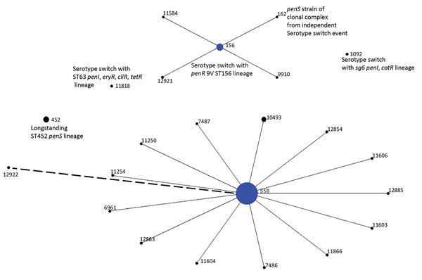
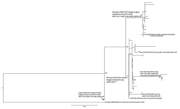
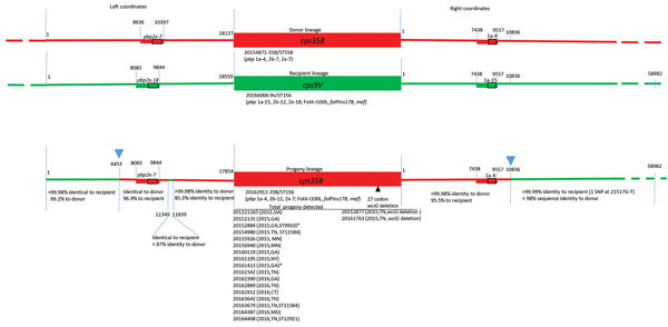
We used whole-genome sequencing to characterize 199 nonvaccine serotype 35B pneumococcal strains that caused invasive pneumococcal disease (IPD) in the United States during 2015-2016 and related these findings to previous serotype 35B IPD data obtained by Active Bacterial Core surveillance. Penicillin-nonsusceptible 35B IPD increased during post-pneumococcal 7-valent conjugate vaccine years (2001-2009) and increased further after implementation of pneumococcal 13-valent conjugate vaccine in 2010. This increase was caused primarily by the 35B/sequence type (ST) 558 lineage. 35B/ST558 and vaccine serotype 9V/ST156 lineages were implicated as cps35B donor and recipient, respectively, for a single capsular switch event that generated emergent 35B/ST156 progeny in 6 states during 2015-2016. Three additional capsular switch 35B variants were identified, 2 of which also involved 35B/ST558 as cps35B donor. Spread of 35B/ST156 is of concern in view of past global predominance of pathogenic ST156 vaccine serotype strains. Protection against serotype 35B should be considered in next-generation pneumococcal vaccines.

Keywords: United States; bacteria; invasive pneumococcal disease; penicillin-binding protein types; pneumococcal 13-valent conjugate vaccine; pneumococcal 7-valent conjugate vaccine; pneumococcal conjugate; pneumococci; recombination; respiratory infections; serotype 35B; serotype expansion; serotype switch; surveillance; switch lineage; vaccines.


Figures

Figure 1
Figure 1
Population snapshot of 199 serotype 35B pneumococcal isolates obtained by ongoing Active Bacterial Core surveillance, United States, 2015–2016, configured by using eBURST (21). Diameters are proportional to number of isolates. Solid lines indicate single-locus variants, and the single dashed line indicates a double-locus variant of ST558. ST, sequence type.
Figure 2
Figure 2
Phylogenetic analysis of potential recipient and serotype switch pneumococcal progeny strains within the ST56 lineage based upon a total alignment of 10,409 core single-nucleotide polymorphisms, United States, 2015–2016. All 20 serotype 35B progeny shown were recovered through Active Bacterial Core surveillance, and all but 2 indicated strains were obtained during 2015–2016. Isolate features are depicted for the 2 major nodes, with exceptions indicated by asterisks within the tree. Bootstrap values are indicated at key nodes. The serotype 9V isolate that was used for the reference recipient sequences described in Figure 3 is indicated as the third isolate from bottom. All isolates above the 9V recipient reference within this major cluster, except where indicated, are also 9V/ST156. Three additional serotype switch ST156 strain types detected by Active Bacterial Core surveillance during 2015–2016 are indicated by asterisks (single isolates of serotypes 31 and 13 and 4 serotype 19A isolates). Scale bar indicates nucleotide substitutions per site.
Figure 3
Figure 3
Diagrammatic representation of cps loci and adjacent regions from donor, recipient and progeny strains depicting serotype switch event for pneumococcal isolates, United States, 2015–2016. Red and green lines in progeny indicate regions of sequence identity or near identity (<2 single-nucleotide polymorphisms/10,000 bp) to the above corresponding donor and recipient sequences, respectively. Rectangles indicate relative locations of PBP gene types for pbp2x and pbp1a. Below each cps locus, a representative reference strain is indicated along with relevant features determined through a bioinformatics pipeline (MLST, PBP type, resistance markers). Junctions between donor and recipient sequences involved in the 2 single recombinational crossovers in the gene replacement event are indicated with blue arrowheads above the progeny diagram, although a single short internal region with sequence identity to the recipient nested within the donor fragment (left coordinates 11349–11839) is also present. Below each green or red segment of the progeny, the level of sequence identity to donor and recipient is provided. The list of each progeny strain, date of isolation, and state is provided. Where MLST is not ST156, its single locus variants (ST9910, ST11584, and ST12921) are included. Two exceptions indicating flanking post-switch recombination within left coordinates 1–6453 are indicated in isolates 20152884 and 20161413 (asterisks): isolate 20152884 had only 99.3%−99.5% identity to recipient and donor over bases 1–3715, and isolate 20161413 had only 99.5%–99.7% identity to recipient and donor over bases 1–2143. Two strains on the right indicate a post-switch deletion event within the wciG putative acetyltransferase gene, which putatively contributes to the acetylation pattern of the serotype 35B polysaccharide (26). MLST, multilocus sequence type; PBP, penicillin-binding protein; ST, sequence type.

Comment in

References

    1. Pilishvili T, Lexau C, Farley MM, Hadler J, Harrison LH, Bennett NM, et al. ; Active Bacterial Core Surveillance/Emerging Infections Program Network. Sustained reductions in invasive pneumococcal disease in the era of conjugate vaccine. J Infect Dis. 2010;201:32–41. 10.1086/648593 - DOI - PubMed
    1. Moore MR, Link-Gelles R, Schaffner W, Lynfield R, Lexau C, Bennett NM, et al. Effect of use of 13-valent pneumococcal conjugate vaccine in children on invasive pneumococcal disease in children and adults in the USA: analysis of multisite, population-based surveillance. Lancet Infect Dis. 2015;15:301–9. 10.1016/S1473-3099(14)71081-3 - DOI - PMC - PubMed
    1. Beall B, McEllistrem MC, Gertz RE Jr, Boxrud DJ, Besser JM, Harrison LH, et al. ; Active Bacterial Core Surveillance/Emerging Infections Program Network. Emergence of a novel penicillin-nonsusceptible, invasive serotype 35B clone of Streptococcus pneumoniae within the United States. J Infect Dis. 2002;186:118–22. 10.1086/341072 - DOI - PubMed
    1. Gertz RE Jr, Li Z, Pimenta FC, Jackson D, Juni BA, Lynfield R, et al. Increased penicillin-nonsusceptibility of nonvaccine serotype (other than 19A and 6A) invasive pneumococci in post 7 valent conjugate vaccine era. J Infect Dis. 2010;201:770–5. 10.1086/650496 - DOI - PubMed
    1. Sharma D, Baughman W, Holst A, Thomas S, Jackson D, da Gloria Carvalho M, et al. Pneumococcal carriage and invasive disease in children before introduction of the 13-valent conjugate vaccine: comparison with the era before 7-valent conjugate vaccine. Pediatr Infect Dis J. 2013;32:e45–53. 10.1097/INF.0b013e3182788fdd - DOI - PubMed