

Red blood cells ageing markers: a multi-parametric analysis
- PMID: 28518051
- PMCID: PMC5448830
- DOI: 10.2450/2017.0318-16
Red blood cells ageing markers: a multi-parametric analysis
Abstract
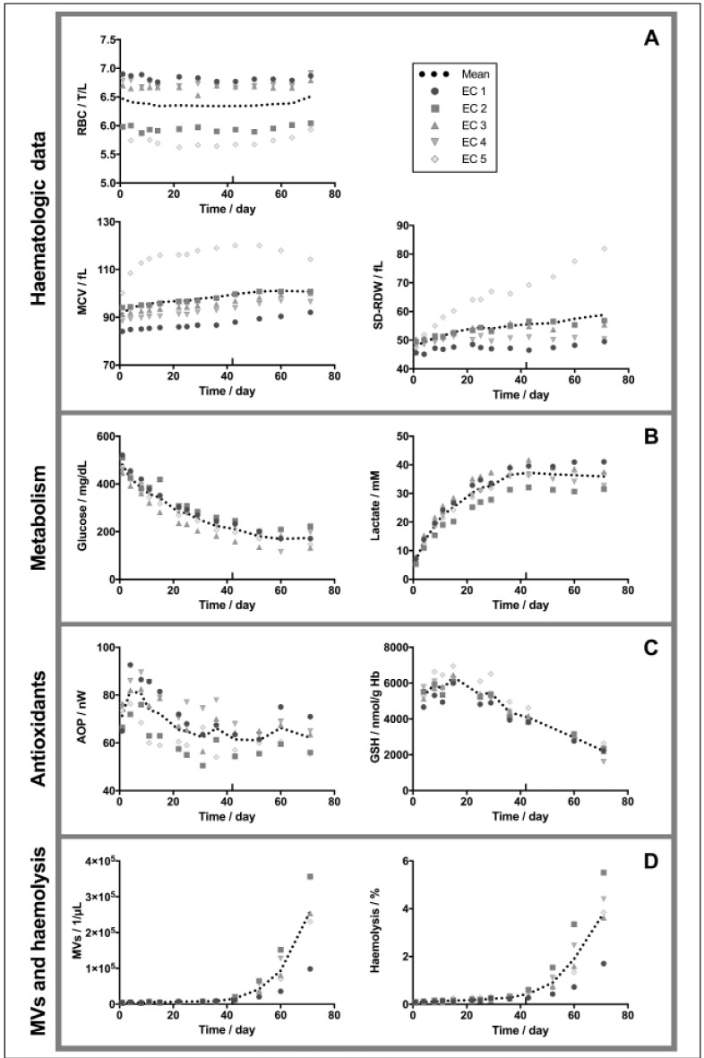
Background: Red blood cells collected in citrate-phosphate-dextrose can be stored for up to 42 days at 4 °C in saline-adenine-glucose-mannitol additive solution. During this controlled, but nevertheless artificial, ex vivo ageing, red blood cells accumulate lesions that can be reversible or irreversible upon transfusion. The aim of the present study is to follow several parameters reflecting cell metabolism, antioxidant defences, morphology and membrane dynamics during storage.
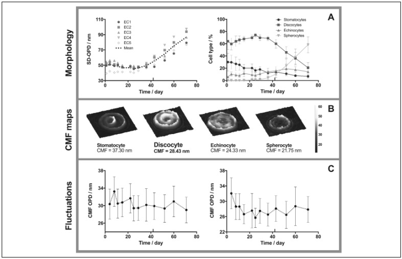
Materials and methods: Five erythrocyte concentrates were followed weekly during 71 days. Extracellular glucose and lactate concentrations, total antioxidant power, as well as reduced and oxidised intracellular glutathione levels were quantified. Microvesiculation, percentage of haemolysis and haematologic parameters were also evaluated. Finally, morphological changes and membrane fluctuations were recorded using label-free digital holographic microscopy.
Results: The antioxidant power as well as the intracellular glutathione concentration first increased, reaching maximal values after one and two weeks, respectively. Irreversible morphological lesions appeared during week 5, where discocytes began to transform into transient echinocytes and finally spherocytes. At the same time, the microvesiculation and haemolysis started to rise exponentially. After six weeks (expiration date), intracellular glutathione was reduced by 25%, reflecting increasing oxidative stress. The membrane fluctuations showed decreased amplitudes during shape transition from discocytes to spherocytes.
Discussion: Various types of lesions accumulated at different chemical and cellular levels during storage, which could impact their in vivo recovery after transfusion. A marked effect was observed after four weeks of storage, which corroborates recent clinical data. The prolonged follow-up period allowed the capture of deep storage lesions. Interestingly, and as previously described, the severity of the changes differed among donors.
Conflict of interest statement
BR also works part-time for Lyncée Tec which commercialises the DHM used in this study. The other Authors declare no conflicts of interest.
Figures
References
MeSH terms
Substances
LinkOut - more resources
Full Text Sources
Other Literature Sources
Molecular Biology Databases
