. 2017 May 18;8(5):e2803.
doi: 10.1038/cddis.2017.210.

Cytoplasmic RAP1 mediates cisplatin resistance of non-small cell lung cancer

Affiliations

Cytoplasmic RAP1 mediates cisplatin resistance of non-small cell lung cancer

Lu Xiao et al. Cell Death Dis. .

Abstract

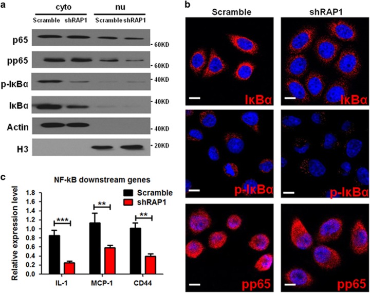
Cytotoxic chemotherapy agents (e.g., cisplatin) are the first-line drugs to treat non-small cell lung cancer (NSCLC) but NSCLC develops resistance to the agent, limiting therapeutic efficacy. Despite many approaches to identifying the underlying mechanism for cisplatin resistance, there remains a lack of effective targets in the population that resist cisplatin treatment. In this study, we sought to investigate the role of cytoplasmic RAP1, a previously identified positive regulator of NF-κB signaling, in the development of cisplatin resistance in NSCLC cells. We found that the expression of cytoplasmic RAP1 was significantly higher in high-grade NSCLC tissues than in low-grade NSCLC; compared with a normal pulmonary epithelial cell line, the A549 NSCLC cells exhibited more cytoplasmic RAP1 expression as well as increased NF-κB activity; cisplatin treatment resulted in a further increase of cytoplasmic RAP1 in A549 cells; overexpression of RAP1 desensitized the A549 cells to cisplatin, and conversely, RAP1 depletion in the NSCLC cells reduced their proliferation and increased their sensitivity to cisplatin, indicating that RAP1 is required for cell growth and has a key mediating role in the development of cisplatin resistance in NSCLC cells. The RAP1-mediated cisplatin resistance was associated with the activation of NF-κB signaling and the upregulation of the antiapoptosis factor BCL-2. Intriguingly, in the small portion of RAP1-depleted cells that survived cisplatin treatment, no induction of NF-κB activity and BCL-2 expression was observed. Furthermore, in established cisplatin-resistant A549 cells, RAP1 depletion caused BCL2 depletion, caspase activation and dramatic lethality to the cells. Hence, our results demonstrate that the cytoplasmic RAP1-NF-κB-BCL2 axis represents a key pathway to cisplatin resistance in NSCLC cells, identifying RAP1 as a marker and a potential therapeutic target for cisplatin resistance of NSCLC.


Conflict of interest statement

The authors declare no conflict of interest.

Figures

Figure 1
Figure 1
Cytoplasmic RAP1 is an indicator of high-grade NSCLC. (a and b) Expression of cytoplasmic and nuclear RAP1 in lung adenocarcinoma (a) and squamous cell carcinoma (b) tissues, as well as peri-tumoral normal tissues (PTNT). (c and d) Expression of cytoplasmic RAP1 in Grade I–III adenocarcinoma (c) and squamous cell carcinoma (d) tissues. (e) Kaplan–Meier analysis and Log-rank (Mantel–Cox) test of the survival between RAP1-high (n=33, expression >0.4) and RAP1-low (n=59, expression <0.4) adenocarcinoma patients. (f) Representative images of RAP1 expression in adenocarcinoma, squamous cell carcinoma and PTNT, arrowhead: cytoplasmic RAP1; scale bar: 10 μm. *P<0.05, **P<0.01, ***P<0.001, Student’s t-test
Figure 2
Figure 2
Proliferation of NSCLC cells is inhibited after Rap1 deletion. (a) RAP1 mRNA expression in 16HBE and HSAEC1-KT (HSA for abbreviation in following) lung epithelial cells and A549, PC9 and HCC827 NSCLC cells measured by qRT-PCR. (b) Western blotting analysis for RAP1 in cytoplasmic and nuclear fractions of lung epithelial and NSCLC cells, representatives of three independent experiments. (c) Immunofluorescence (IF) detection of RAP1 (red) in 16HBE and NSCLC cells. Arrowhead: cytoplasmic RAP1; scale bar: 5 μm. (d) RAP1 mRNA expression in A549, PC9 and HCC827 cells after transducing lentiviral vector coding two shRNAs against RAP1 (shRAP1) or scramble shRNA. (e) Proliferation of NSCLC cells after transduction with shRAP1 or scramble shRNA. Cells were plated in 96-well plates, cultured for 96 h and viability determined through cell counting kit-8 (CCK-8) assay. (f) Colony-formation assays for A549 cells after transduction with shRAP1 or scramble shRNA, detected by Giemsa staining. Statistics were generated from three independent experiments. **P<0.01 ***P<0.001, Student’s t-test (two groups) or one-way analysis of variance with Bonferroni’s correction (three or more groups); error bar: ±S.D.
Figure 3
Figure 3
RAP1 acts through NF-κB signaling to support NSCLC cell growth. (a and b) Western blotting analysis (a) and IF assay (b) of NF-κB signaling factors: p65, phosphorylated-p65 (pp65), IκBα, and phosphorylated-IκBα (p-IκBα) in the cytoplasmic and nuclear fractions of A549 cells transduced with shRAP1 or scramble shRNA. Scale bar: 5 μm. (c) Expression of genes positively regulated by NF-κB signaling in A549 cells transduced with shRAP1 or scramble shRNA. Statistics were generated from three independent experiments. **P<0.01, ***P<0.001, Student’s t-test; error bar: ±S.D.
Figure 4
Figure 4
RAP1 desensitizes NSCLC cells to CP-induced cell death. (a) shRAP1- or scramble shRNA-transduced A549 cells were treated with different concentrations of CP for 48 h, and cell viability was determined with CCK-8 assay and normalized to the same type of cells cultured without CP. (b) Proliferation of A549 cells after transduction with control (CTRL) or RAP1-overexpressing (RAP1) lentiviral vectors. (c) Cell viability of A549 cells transduced with RAP1 or CTRL viruses and treated with different concentrations of CP for 48 h, after normalization to the same type of cells cultured in CP-free media. (d) Competition assay of the untransduced (GFP) cells co-cultured with the cells transduced with the indicated different RAP1 modification lentiviruses (GFP+). Cells were treated with 2 μM of CP and harvested at 24 and 72 h. Percentage of GFP+ cells were analyzed for the viable population determined by 4,6-diamidino-2-phenylindole staining. (e and f) Untransduced (UnTxD) A549 cells and A549 cells with RAP1 overexpression (RAP1) or RAP1 knockdown (shRAP1) were cultured in media containing 1 μM CP (+) or CP-free media (−) for 24 h and analyzed for the apoptotic status with western blotting analysis for cleaved caspase-3 (C-Cas3) and BCL-2 (e) or with flow cytometric analysis of Annexin-V (f), representatives of three independent experiments. Statistics were generated from three independent experiments. *P<0.05, **P<0.01, ***P<0.001, Student’s t-test; error bar: ±S.D.
Figure 5
Figure 5
CP resistance is associated with RAP1-dependent NF-κB signaling induction. (a) Schema of stepwise CP treatment on A549 cells. (b and c) RAP1 expression and NF-κB activation during CP treatment. A549 cells treated with CP as depicted in panel (a) were harvested at the indicated time points; the protein expression of RAP1, p65, pp65 and p-IκBα in the cytoplasmic and nuclear fractions was measured with western blotting analyses (b), representatives of three independent experiments; and the mRNA expression of NF-κB signaling downstream factors was analyzed with qRT-PCR (c). (d) A549 cells with overexpressed RAP1 were treated with CP as depicted in panel a. RAP1, pp65 and p-IκBα were detected in the cytoplasmic (Cyto) and nuclear (Nu) fractions of cell lysate at the indicated time points, representatives of three independent experiments. (e) Cell viability of A549 cells transduced with shRAP1 or scramble shRNA at different time points during the sequential CP treatment as depicted in panel a, normalized to the same type of cells cultured in CP-free media in respective time point. (f) Nuclear (upper panel) and cytoplasmic (lower panel) fractions were isolated from A549 cells transduced with shRAP1 or scramble shRNA and treated with 0.5 μM of CP for 24 h and assayed for p65, pp65 and p-IκBα protein levels, representatives of three independent experiments. (g) mRNA expression of NF-κB signaling downstream factors in shRAP1-transduced A549 cells after 24 h culture in the presence or absence of 0.5 μM of CP. Statistics were generated from three independent experiments. **P<0.01, ***P<0.001, Student’s t-test; error bar: ±S.D.
Figure 6
Figure 6
RAP1 deletion is lethal to CP-resistant NSCLC cells. (a and b) Protein expression of RAP1, pp65, p-IκBα (a) and mRNA expression of NF-κB signaling downstream factors (b) in control (CTRL), RAP1-overexpressed (RAP1) and CP-resistant (A549R) cells, representatives of three independent experiments. (c) Cell proliferation of A549R cells transduced with scramble shRNA or shRAP1 assayed with CCK-8 staining at the time point of transduction. Cells were precultured in CP-free media for 24 h before transduction. (d) RAP1-deleted A549 cells and scramble shRNA-transduced A549R cells were cultured in media containing 1 μM CP (+) or CP-free media (−) for 24 h before being lysed to harvest protein. Lentiviral shRAP1 was introduced to A549R cells precultured in CP-free media for 24 h and protein harvested 12 h after transduction. Cleaved caspase-3 and BCL-2 were detected by western blotting. (e) IF assay of cleaved caspase-3 (red) in untreated A549 and A549R cells transduced with scramble shRNA or shRAP1. (f) Flow cytometric analysis for Annexin-V staining in A549 and A549R cells after RAP1 deletion. A549 cells were maintained in CP-free media and A549R cells were preconditioned in CP-free media for 24 h before analysis. Statistics were generated from three independent experiments. *P<0.05, **P<0.01, ***P<0.001, Student’s t-test; error bar: ±S.D.
Figure 7
Figure 7
Schematic depiction of the proposed model. CP generates DNA damage, which eventually leads to cell apoptosis. Meanwhile, RAP1 is upregulated after CP treatment, possibly through a direct or indirect induction by DNA damage response. The cytoplasmic fraction of RAP1 thus acts to facilitate the IKK-mediated activation of NF-κB signaling, which subsequently induces transcription of downstream factors, including the apoptosis inhibitor BCL-2. Therefore, RAP1 functions through activating NF-κB signaling to mediate resistance to CP

References

    1. Gridelli C, Rossi A, Carbone DP, Guarize J, Karachaliou N, Mok T et al. Non-small-cell lung cancer. Nat Rev Dis Primers 2015; 1: 15009. - PubMed
    1. Schaake-Koning C, van den Bogaert W, Dalesio O, Festen J, Hoogenhout J, van Houtte P et al. Effects of concomitant Cisplatin and radiotherapy on inoperable non-small-cell lung cancer. N Engl J Med 1992; 326: 524–530. - PubMed
    1. Olaussen KA, Dunant A, Fouret P, Brambilla E, Andre F, Haddad V et al. DNA repair by ERCC1 in non-small-cell lung cancer and Cisplatin-based adjuvant chemotherapy. N Engl J Med 2006; 355: 983–991. - PubMed
    1. Mitsudomi T, Morita S, Yatabe Y, Negoro S, Okamoto I, Tsurutani J et al. Gefitinib versus Cisplatin plus docetaxel in patients with non-small-cell lung cancer harbouring mutations of the epidermal growth factor receptor (WJTOG3405): an open label, randomised phase 3 trial. Lancet Oncol 2010; 11: 121–128. - PubMed
    1. Scagliotti GV, Parikh P, von Pawel J, Biesma B, Vansteenkiste J, Manegold C et al. Phase III study comparing Cisplatin plus gemcitabine with Cisplatin plus pemetrexed in chemotherapy-naive patients with advanced-stage non-small-cell lung cancer. J Clin Oncol 2008; 26: 3543–3551. - PubMed