Analysis of heterogeneity in T2-weighted MR images can differentiate pseudoprogression from progression in glioblastoma
- PMID: 28520730
- PMCID: PMC5435159
- DOI: 10.1371/journal.pone.0176528
Analysis of heterogeneity in T2-weighted MR images can differentiate pseudoprogression from progression in glioblastoma
Abstract
Purpose: To develop an image analysis technique that distinguishes pseudoprogression from true progression by analyzing tumour heterogeneity in T2-weighted images using topological descriptors of image heterogeneity called Minkowski functionals (MFs).
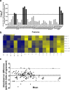
Methods: Using a retrospective patient cohort (n = 50), and blinded to treatment response outcome, unsupervised feature estimation was performed to investigate MFs for the presence of outliers, potential confounders, and sensitivity to treatment response. The progression and pseudoprogression groups were then unblinded and supervised feature selection was performed using MFs, size and signal intensity features. A support vector machine model was obtained and evaluated using a prospective test cohort.
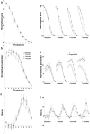
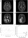
Results: The model gave a classification accuracy, using a combination of MFs and size features, of more than 85% in both retrospective and prospective datasets. A different feature selection method (Random Forest) and classifier (Lasso) gave the same results. Although not apparent to the reporting radiologist, the T2-weighted hyperintensity phenotype of those patients with progression was heterogeneous, large and frond-like when compared to those with pseudoprogression.
Conclusion: Analysis of heterogeneity, in T2-weighted MR images, which are acquired routinely in the clinic, has the potential to detect an earlier treatment response allowing an early change in treatment strategy. Prospective validation of this technique in larger datasets is required.
Conflict of interest statement
Figures


Similar articles
-
Differentiation of Pseudoprogression from True Progressionin Glioblastoma Patients after Standard Treatment: A Machine Learning Strategy Combinedwith Radiomics Features from T1-weighted Contrast-enhanced Imaging.BMC Med Imaging. 2021 Feb 3;21(1):17. doi: 10.1186/s12880-020-00545-5. BMC Med Imaging. 2021. PMID: 33535988 Free PMC article.
-
Machine learning-based radiomic evaluation of treatment response prediction in glioblastoma.Clin Radiol. 2021 Aug;76(8):628.e17-628.e27. doi: 10.1016/j.crad.2021.03.019. Epub 2021 May 1. Clin Radiol. 2021. PMID: 33941364
-
Prediction of pseudoprogression in post-treatment glioblastoma using dynamic susceptibility contrast-derived oxygenation and microvascular transit time heterogeneity measures.Eur Radiol. 2024 May;34(5):3061-3073. doi: 10.1007/s00330-023-10324-9. Epub 2023 Oct 18. Eur Radiol. 2024. PMID: 37848773
-
Pseudoprogression versus true progression in glioblastoma patients: A multiapproach literature review. Part 2 - Radiological features and metric markers.Crit Rev Oncol Hematol. 2021 Mar;159:103230. doi: 10.1016/j.critrevonc.2021.103230. Epub 2021 Jan 27. Crit Rev Oncol Hematol. 2021. PMID: 33515701 Review.
-
Pseudoprogression versus true progression in glioblastoma patients: A multiapproach literature review: Part 1 - Molecular, morphological and clinical features.Crit Rev Oncol Hematol. 2021 Jan;157:103188. doi: 10.1016/j.critrevonc.2020.103188. Epub 2020 Dec 8. Crit Rev Oncol Hematol. 2021. PMID: 33307200 Review.
Cited by
-
Machine learning and glioma imaging biomarkers.Clin Radiol. 2020 Jan;75(1):20-32. doi: 10.1016/j.crad.2019.07.001. Epub 2019 Jul 29. Clin Radiol. 2020. PMID: 31371027 Free PMC article. Review.
-
Effective Detection and Monitoring of Glioma Using [18F]FPIA PET Imaging.Biomedicines. 2021 Jul 13;9(7):811. doi: 10.3390/biomedicines9070811. Biomedicines. 2021. PMID: 34356874 Free PMC article.
-
Perfusion MRI in treatment evaluation of glioblastomas: Clinical relevance of current and future techniques.J Magn Reson Imaging. 2019 Jan;49(1):11-22. doi: 10.1002/jmri.26306. J Magn Reson Imaging. 2019. PMID: 30561164 Free PMC article. Review.
-
Artificial intelligence in the radiomic analysis of glioblastomas: A review, taxonomy, and perspective.Front Oncol. 2022 Aug 2;12:924245. doi: 10.3389/fonc.2022.924245. eCollection 2022. Front Oncol. 2022. PMID: 35982952 Free PMC article. Review.
-
Unraveling response to temozolomide in preclinical GL261 glioblastoma with MRI/MRSI using radiomics and signal source extraction.Sci Rep. 2020 Nov 12;10(1):19699. doi: 10.1038/s41598-020-76686-y. Sci Rep. 2020. PMID: 33184423 Free PMC article.
References
-
- Michelson K, De Raedt H. Integral-geometry morphological image analysis. Phys Rep 2001; 347:461–538.
MeSH terms
Grants and funding
LinkOut - more resources
Full Text Sources
Other Literature Sources
Medical

