. 2017 Jun 20;45(11):6259-6264.
doi: 10.1093/nar/gkx446.

Mass spectrometry for serine ADP-ribosylation? Think o-glycosylation!

Affiliations

Mass spectrometry for serine ADP-ribosylation? Think o-glycosylation!

Juan J Bonfiglio et al. Nucleic Acids Res. .

Abstract

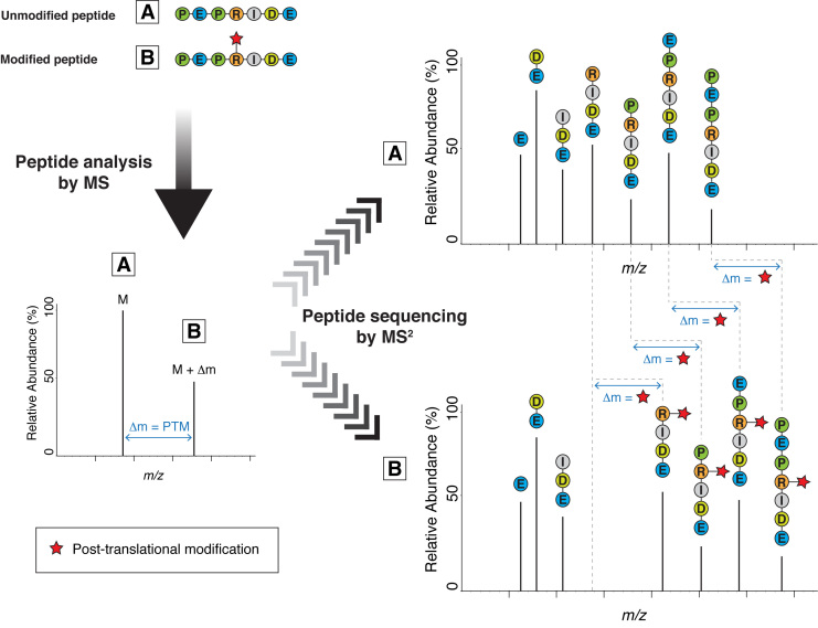
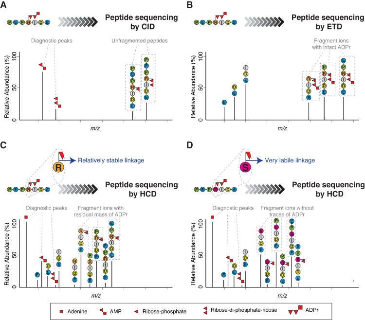
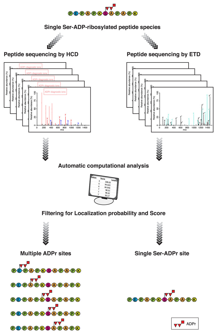
Protein ADP-ribosylation (ADPr), a biologically and clinically important post-translational modification, exerts its functions by targeting a variety of different amino acids. Its repertoire recently expanded to include serine ADPr, which is emerging as an important and widespread signal in the DNA damage response. Chemically, serine ADPr (and more generally o-glycosidic ADPr) is a form of o-glycosylation, and its extreme lability renders it practically invisible to standard mass spectrometry approaches, often leading to erroneous localizations. The knowledge from the mature field of o-glycosation and our own initial difficulties with mass spectrometric analyzes of serine ADPr suggest how to avoid these misidentifications and fully explore the scope of o-glycosidic ADPr in DNA damage response and beyond.


Figures

Figure 1.
Figure 1.
Mass spectrometry (MS) for the analysis of post-translational modifications (PTMs) via the identification of the modified peptides. When we analyze peptides by mass spectrometry, we first acquire an MS spectrum (left-hand spectrum) to determine the molecular mass of the peptide molecules entering the spectrometer (the ‘precursors’). Next, each of these peptide species is selected and fragmented into smaller pieces, producing a set of fragment ions (right-hand spectra). From these fragment ions (MS2 peaks), we can extract information about the sequence of the peptide. The masses of adjacent fragment ions differ by the masses of the corresponding amino acids. The presence of a typical PTM changes the mass of the peptide, the modified amino acid residue and all fragment ions containing the modified residue by Δm.
Figure 2.
Figure 2.
Different fragmentation methods applied to ADP-ribosylated peptides. (A) Schematic representation of a typical collision-induced dissociation (CID) fragmentation spectrum from an ADP-ribosylated peptide. The CID spectrum contains more ions formed through fragmentation of the ADP-ribose itself than through fragmentation of the peptide backbone. As a consequence, it yields little or no information about the peptide sequence. However, CID can be used to confirm the presence of ADP-ribose on a precursor by generating modification-specific diagnostic peaks (e.g. AMP). (B) Schematic representation of a typical electron-transfer dissociation (ETD) fragmentation spectrum from an ADP-ribosylated peptide. The ETD spectrum is rich in information about the peptide sequence and the site of modification. A characteristic feature of ETD is the retention of the intact ADP-ribose. The masses of adjacent fragment ions differ by the masses of the corresponding amino acids. The presence of an ADP-ribose changes the mass of the modified amino acid residue and the ions containing the modified residue by 541.06 Da. (C) Schematic representation of a typical higher-energy collisional dissociation (HCD) fragmentation spectrum from an ADP-ribosylated peptide in which the ADP-ribose linkage to the modified amino acid is relatively stable under HCD fragmentation (e.g. ADPr on Arg). The HCD spectrum contains ions from fragmentation of both the peptide backbone and ADP-ribose. Usually, the HCD spectrum is dominated by one of the diagnostic ions generated by the internal fragmentation of ADP-ribose (adenine, at m/z 136.06), but additional fragments are also detected corresponding to the peptide with a residual part of ADP-ribose attached (typically ribose phosphate-H2O). These fragment ions provide the basis for determining the site of modification. (D) Schematic representation of a typical HCD spectrum from an ADP-ribosylated peptide in which the ADP-ribose linkage to the modified amino acid is very labile under HCD fragmentation (e.g. ADPr on Ser). The HCD spectrum contains ions from both the peptide backbone and from the ADP-ribose fragmentation. This spectrum can contain peptide fragment ions carrying part of ADP-ribose, but the strongest ion series is not that of a modified sequence, but of the native identified peptide. These de-modified fragment ions contain no information for determining the modification site. Since search algorithms score these de-modified ions as unmodified ions, conventional analysis of serine ADPr by HCD can lead to multiple, erroneous localizations arising from variations in spectrum quality. See Figure 3.
Figure 3.
Figure 3.
Conventional analysis of serine ADPr by HCD can lead to multiple and erroneous ADPr site assignments. Schematic representation of a standard MS workflow illustrating the problematic behavior of Ser-ADP-ribosylated peptides under HCD fragmentation. In this representation, the same single Ser-ADP-ribosylated peptide species is subjected to MS analysis either using HCD or ETD fragmentation (left and right-hand respectively). The acquired mass spectra are searched against the appropriate database using any one of a number of database-search systems (e.g. Maxquant, Mascot, etc). The initial list of ADPr sites obtained after the automatic computational analysis is filtered by different criteria to eliminate unreliable (low scoring) or ambiguous (low localization score) results. The final filtered list of ADPr sites obtained from ETD fragmentation contains the same unique ADPr site as the single ADP-ribosylated peptide species initially subjected to MS analysis (bottom right-hand). On the other hand, the applying of the same filtering parameters to the sites found from HCD fragmentation data, reveals fictional ADPr sites in the final list, illustrating the fundamental weakness of this analysis approach (bottom left-hand). Readers can evaluate the misleading localization results provided by HCD MS spectra by re-analyzing publicly available data for themselves. A dataset containing HCD and ETD spectra from the same sample is recommended to demonstrate the fundamental weakness of HCD as well as the necessity of ETD for accurate localization of Ser-ADPr (31,37). Alternatively, large published HCD datasets from ADP-ribosylated peptides can also be re-analyzed permitting modification of chemically impossible ‘decoy’ sites (like alanine) to illustrate the weakness of automatic computational analysis of HCD data. This form of analysis illustrates the relative influence of data and prior assumptions on results.

References

    1. Hedberg C., Itzen A.. Molecular perspectives on protein adenylylation. Acs. Chem. Biol. 2015; 10:12–21. - PubMed
    1. Feng F.Y., de Bono J.S., Rubin M.A., Knudsen K.E.. Chromatin to clinic: the molecular rationale for PARP1 inhibitor function. Mol. Cell. 2015; 58:925–934. - PMC - PubMed
    1. Bock F.J., Chang P.. New directions in PARP biology. FEBS J. 2016; 283:4017–4031. - PubMed
    1. Gupte R., Liu Z., Kraus W.L.. PARPs and ADP-ribosylation: recent advances linking molecular functions to biological outcomes. Genes Dev. 2017; 31:101–126. - PMC - PubMed
    1. Martin-Hernandez K., Rodriguez-Vargas J.M., Schreiber V., Dantzer F.. Expanding functions of ADP-ribosylation in the maintenance of genome integrity. Semin. Cell Dev. Biol. 2017; 63:92–101. - PubMed

LinkOut - more resources