. 2017 Jun 20;45(11):6628-6643.
doi: 10.1093/nar/gkx426.

The DEAD-box helicase Mss116 plays distinct roles in mitochondrial ribogenesis and mRNA-specific translation

Affiliations

The DEAD-box helicase Mss116 plays distinct roles in mitochondrial ribogenesis and mRNA-specific translation

Dasmanthie De Silva et al. Nucleic Acids Res. .

Abstract

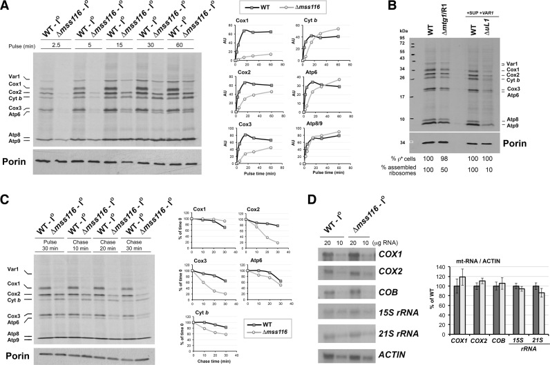
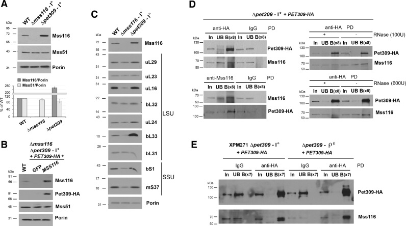
Members of the DEAD-box family are often multifunctional proteins involved in several RNA transactions. Among them, yeast Saccharomyces cerevisiae Mss116 participates in mitochondrial intron splicing and, under cold stress, also in mitochondrial transcription elongation. Here, we show that Mss116 interacts with the mitoribosome assembly factor Mrh4, is required for efficient mitoribosome biogenesis, and consequently, maintenance of the overall mitochondrial protein synthesis rate. Additionally, Mss116 is required for efficient COX1 mRNA translation initiation and elongation. Mss116 interacts with a COX1 mRNA-specific translational activator, the pentatricopeptide repeat protein Pet309. In the absence of Mss116, Pet309 is virtually absent, and although mitoribosome loading onto COX1 mRNA can occur, activation of COX1 mRNA translation is impaired. Mutations abolishing the helicase activity of Mss116 do not prevent the interaction of Mss116 with Pet309 but also do not allow COX1 mRNA translation. We propose that Pet309 acts as an adaptor protein for Mss116 action on the COX1 mRNA 5΄-UTR to promote efficient Cox1 synthesis. Overall, we conclude that the different functions of Mss116 in the biogenesis and functioning of the mitochondrial translation machinery depend on Mss116 interplay with its protein cofactors.


Figures

Figure 1.
Figure 1.
Mss116 is essential for OXPHOS system assembly and function. See also Supplementary Figure S1 and Table S1. (A) Growth test using serial dilutions of the indicated strains in complete fermentable (YPD) and respiratory (YEPG) media. Pictures were taken after 2 and 4 days of growth at 30°C. (B) Total mitochondrial cytochrome spectra from the WT and Δmss116 strains. (C) Assessment of the respiration and OXPHOS enzyme functions. Polarographic determination of endogenous cell respiration, and oxidation of NADH or Ascorbate-TMPD in isolated mitochondria. Spectrophotometric measurement of NCCR (NADH cytochrome c reductase), QCCR (Ubiquinol cytochrome c reductase, cytochrome c oxidase (COX) and oligomycin-sensitive ATPase. The bars indicate the means ± SD from at least three independent sets of measurements. (D) Steady-state levels of the indicated proteins estimated by immunoblot analyses of mitochondrial proteins. The Immunoblot for Cox1 is presented in duplicate, representing two exposure times. (E) Immunoblot analysis of OXPHOS complexes extracted from mitochondria using native conditions and separated by blue native (BN)-polyacrylamide gel electrophoresis.
Figure 2.
Figure 2.
Mss116 interacts with the mitoribosomal LSU and its absence affects mitoribosome biogenesis. (A and B) Sucrose gradient sedimentation analyses of mitoribosomal proteins and assembly factors on mitochondrial extracts prepared from the WT and Δmss116 strains in the presence of 0.8% Triton X-100 and the conditions stated including either Mg2+ (A) or ethylenediaminetetraacetic acid (EDTA) (B). (C) Steady-state levels of mitoribosomal proteins estimated by immunoblot analyses. Porin was used as a loading control. (D) Sucrose gradient sedimentation analyses of SSU and LSU in mitochondrial extracts from the indicated strains prepared in the presence of 5 mM EDTA. The fractions were used to measure total RNA concentration (bottom) and to analyze the distribution of Mss116 and the ribosomal proteins by immunoblotting (top).
Figure 3.
Figure 3.
Mitochondrial protein synthesis is altered in the absence of Mss116. (A) In vivo mitochondrial protein synthesis in the indicated strains following incorporation of [35S]-methionine into newly synthesized polypeptides as a function of time. Proteins were separated by sodium dodecyl sulphate-polyacrylamide gel electrophoresis (SDS-PAGE), transferred to a nitrocellulose membrane and exposed to X-ray film. The graphs show the densitometry values obtained by using the histogram function of the Adobe Photoshop program on digitalized images. The values were normalized by the immunoblot signal of Porin and expressed relative to the control. Two independent repetitions did not differ by more than 5%. (B) In vivo mitochondrial protein synthesis as in panel (A), during 15 min, using the indicated strains. Immunoblotting for Porin was used as a loading control. Two mitoribosomal mutants were used. The strain Δmtg1/R1 carries a null allele of the mtLSU assembly factor MTG1 and a partial suppressor mutation in the 21S rRNA. The strain ΔuL1 carries a null allele of the mtLSU subunit uL1. For each mutant, the corresponding wild-type (WT) control strain was used. In all cases, the percentage of cells carrying mitochondrial DNA (ρ+) was near 100%. (C) Pulse-chase mitochondrial translation. Following a 30-min pulse, cold methionine and puromycin were added and the stability of newly-synthesized mitochondrial proteins was followed as a function of time at 30°C. Signals were quantified as in (A) and expressed as percentage of chase time zero. (D) Northern blot analyses of total RNA probed for COX1, COX2, COB and 15S and 21S rRNA. After processing, the membranes were exposed to X-ray film, and quantification of the signals was carried out as in panel (A). The values were normalized by the signal of ACT1 mRNA as the loading control and expressed relative to the control. Error bars represent the mean ± SD of three independent repetitions. *P < 0.05.
Figure 4.
Figure 4.
Mss116 is required for COX1 mRNA translation initiation and elongation. See also Supplementary Figures S2 and 3. (A) Scheme depicting the mitochondrial genotype of strains carrying mitochondrial ARG8 as a reporter of mRNA translation. (B) In vivo mitochondrial protein synthesis in the presence of cycloheximide using the strains presented in panel (A). Porin was used as a loading control. (C) Growth test using serial dilutions of the indicated strains in minimum media supplemented or not with arginine and in complete respiratory medium (YEPG). Pictures were taken after 2 days of growth at 30°C.
Figure 5.
Figure 5.
Mss116 physically interacts with Pet309 and support its stability. See also Supplementary Figure S4. (A–C) Steady-state levels of (A) Mss116 and Mss51, (B) Mss116, Mss51 and HA-tagged Pet309, and (C) mitoribosomal proteins. In (B), a plasmid expressing GFP was used as a control. Mitochondrial proteins were separated by SDS-PAGE and analyzed by immunoblotting. Antibody–protein complexes were visualized by chemiluminescence and exposition to an X-ray film. The graph in panel (A) shows the densitometry values obtained by using the histogram function of the Adobe Photoshop program on digitalized images. The values were normalized by the signal of Porin as the loading control and are expressed relative to the control. Bars represent the average of three independent experiments ± SD. (D and E) Immunoprecipitation of Mss116 or Pet309-HA from mitochondrial extracts prepared from the indicated strains, using HA-conjugated magnetic beads. In (D), the extracts where either left untreated (left panel) or incubated with the indicated concentrations of RNase A for 30 min at 4°C (right panel). In, input; UB, unbound; B, bound. The concentration of B is 8-fold higher (x8) or 7-fold higher (x7) than that of UB.
Figure 6.
Figure 6.
Mitoribosome loading onto COX1 mRNA can occur in the absence of Mss116, but the COX1 mRNA is not detected in polysomes. (A) Scheme depicting the experimental protocol for mito-polysome profiling. (B–D) Sucrose gradient sedimentation analyses of mitoribosomal subunits, monosomes and polysomes in extracts obtained from mitochondria isolated from the indicated strains, all (including the WT strain) carrying the mtDNA SUP- and ectopic VAR1 expressing plasmids. In each case, the upper panel is an immunoblot analysis of mitoribosome subunit markers. The Immunoblot for fractions 16–28 is presented in duplicate, with short (as for the blot with fractions 1–15) or longer exposure time. The graphs in the lower panels represent the relative amount of the indicated rRNAs and mRNAs in the gradient fraction, estimated by qPCR. (E) Relative mRNA loading into the mt-SSU, mitoribosomes and polysomes estimated using samples from sucrose gradient fractions in panels (B–D). For each strain, the relative mRNA percentage in each structure was calculated. The bars represent the average of three independent experiments ± SD.
Figure 7.
Figure 7.
The catalytic activity of Ms116 is required for its role in COX1 mRNA translation. (A) Diagram of the Mss116 protein depicting the conserved core with nine motifs characteristic of the DEAD-box subfamily of DExH/D-box proteins (black bars). Asterisks indicate the locations of Mss116 mutations analyzed in this work. (B) Steady-state levels of Mss116 and HA-tagged Pet309 in WT and Δmss116 and Δmss116Δpet309+PET309-HA strains expressing GFP as a negative control, WT MSS116 or the indicated mss116 variants. Porin was used as the loading control. (C) Immunoprecipitation of Pet309-HA from mitochondrial extracts obtained from the indicated strains, using HA-conjugated magnetic beads. Mss116 was also detected in the different fractions. In, input; UB, unbound; B, bound. (D) In vivo mitochondrial protein synthesis in the WT and Δmss116 (Δ) strains expressing GFP as a negative control, WT MSS116 or the indicated mss116 variants, following the incorporation of 35S-methionine into newly synthesized proteins (30-min pulse) in the presence of cycloheximide to inhibit cytoplasmic protein synthesis. Immunoblot for Porin was used as a loading control.
Figure 8.
Figure 8.
Roles of Mss116 in mitoribogenesis and COX1 mRNA translation. (A) Primary nucleotide sequence of the COX1 5΄-UTR with the two in silico-predicted Pet309-binding sites shaded in cyan, and the AUG translational start codon and downstream sequence in bold. (B) Model of the secondary structure of COX1 mRNA 5΄UTR. The mfold-quikfold program for RNA folding prediction based on thermodynamic parameters (56) was used to analyze the folding with minimum free energy (ΔG) of the COX1 mRNA 5΄UTR. The potential Pet309 binding sites from (A) in the COX1 mRNA 5΄UTR are shaded in blue. The broad Mss51 interaction site, previously determined by Y3H experiments (45) is indicated. (C) Schematics portraying the distinct roles of Mss116 in mitoribosome assembly and COX1 mRNA translation. To model the interaction between Mss116 and Pet309 we performed a docking analysis using the server GrammX (69). We used the crystallographic model of Mss116 co-purified with ssRNA (PDB: 4TZ6) (70), and a model of Pet309 obtained from the ITASSER server (71).

Similar articles

Cited by

References

    1. De Silva D., Tu Y.T., Amunts A., Fontanesi F., Barrientos A.. Mitochondrial ribosome assembly in health and disease. Cell Cycle. 2015; 14:2226–2250. - PMC - PubMed
    1. Amunts A., Brown A., Bai X., Llácer J.L., Hussain T., Emsley P., Long F., Murshudov G., Scheres S.H.W., Ramakrishnan V.. Structure of the yeast mitochondrial large ribosomal subunit. Science. 2014; 343:1485–1489. - PMC - PubMed
    1. Amunts A., Brown A., Toots J., Scheres S.H.W., Ramakrishnan V.. The structure of the human mitochondrial ribosome. Science. 2015; 348:95–98. - PMC - PubMed
    1. Greber B.J., Bieri P., Leibundgut M., Leitner A., Aebersold R., Boehringer D., Ban N.. Ribosome. The complete structure of the 55S mammalian mitochondrial ribosome. Science. 2015; 348:303–308. - PubMed
    1. Desai N., Brown A., Amunts A., Ramakrishnan V.. The structure of the yeast mitochondrial ribosome. Science. 2017; 355:528–531. - PMC - PubMed

MeSH terms

LinkOut - more resources