Review
. 2017 Nov 1;109(11):djx059.
doi: 10.1093/jnci/djx059.

Drugging the Cancers Addicted to DNA Repair

Affiliations
Review

Drugging the Cancers Addicted to DNA Repair

Jac A Nickoloff et al. J Natl Cancer Inst. .

Abstract

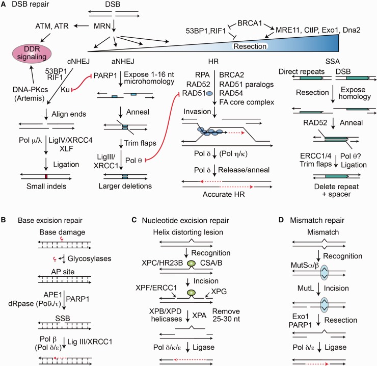
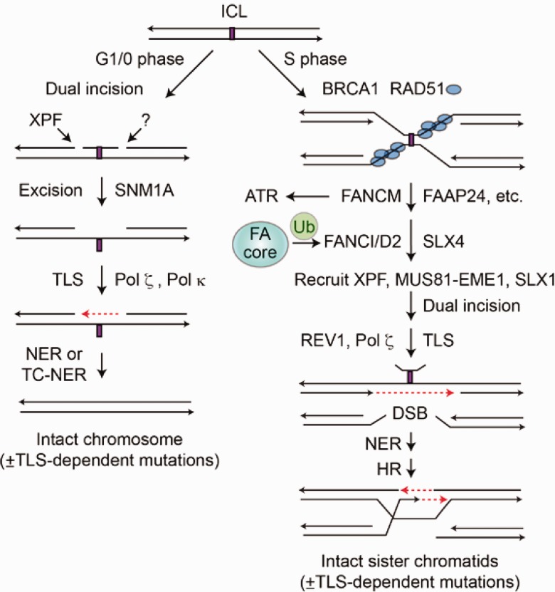
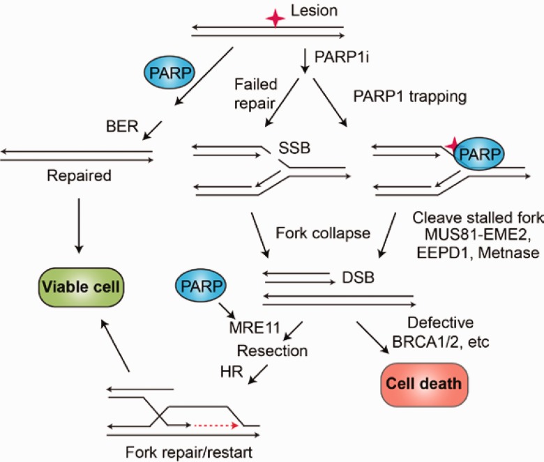
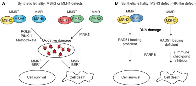
Defects in DNA repair can result in oncogenic genomic instability. Cancers occurring from DNA repair defects were once thought to be limited to rare inherited mutations (such as BRCA1 or 2). It now appears that a clinically significant fraction of cancers have acquired DNA repair defects. DNA repair pathways operate in related networks, and cancers arising from loss of one DNA repair component typically become addicted to other repair pathways to survive and proliferate. Drug inhibition of the rescue repair pathway prevents the repair-deficient cancer cell from replicating, causing apoptosis (termed synthetic lethality). However, the selective pressure of inhibiting the rescue repair pathway can generate further mutations that confer resistance to the synthetic lethal drugs. Many such drugs currently in clinical use inhibit PARP1, a repair component to which cancers arising from inherited BRCA1 or 2 mutations become addicted. It is now clear that drugs inducing synthetic lethality may also be therapeutic in cancers with acquired DNA repair defects, which would markedly broaden their applicability beyond treatment of cancers with inherited DNA repair defects. Here we review how each DNA repair pathway can be attacked therapeutically and evaluate DNA repair components as potential drug targets to induce synthetic lethality. Clinical use of drugs targeting DNA repair will markedly increase when functional and genetic loss of repair components are consistently identified. In addition, future therapies will exploit artificial synthetic lethality, where complementary DNA repair pathways are targeted simultaneously in cancers without DNA repair defects.


Figures

Figure 1.
Figure 1.
DNA repair pathways in mammalian cells. A) Double-strand breaks (DSBs) activate DNA damage response signaling including checkpoint arrest through ATM, ATR, and DNA-PKcs. DSB repair pathway choice is determined by the amount of 5’ end resection at the DSB, inhibited by 53BP1/RIF1, promoted by BRCA1/CtIP. MRE11 initiates limited end resection, and this is followed by Exo1/EEPD1 and Dna2 nucleases for extensive resection. 53BP1/RIF1 and Ku protect DSB ends from resection, promoting classical nonhomologous end joining (cNHEJ). PARP1 competes with Ku and promotes limited end resection for alternative nonhomologous end joining (aNHEJ). RAD51 catalyzes invasion by the resected 3' end into the sister or other homologous sequences, and Pol δ catalyzes repair synthesis across the DSB. The amount of 3’ end resection regulates DSB pathway choice. cNHEJ requires little or no end resection, aNHEJ requires limited resection, and homologous recombination (HR) and single-strand annealing (SSA) require extensive resection. DNA polymerase θ (Pol θ) promotes a microhomology search by the opposing 3’ single strands after short resection in aNHEJ. The HR subpathway SSA requires extensive end resection to expose large homologous regions, usually direct repeats, with RAD52 promoting annealing, producing large deletions between direct repeats. B) Base excision repair repairs minor lesions (eg, oxidized bases), promoted by PARP1. Repair intermediates include an abasic site and nicking of the DNA backbone, with short gap filling by Pol β. C) Nucleotide excision repair (NER) repairs large helix-distorting lesions and involves excision of about 15 nucleotides on either side of the lesion, followed by gap filling. GG-NER and TC-NER differ in lesion recognition by XPA or CSA/B, respectively. D) Mismatch repair involves recognition of the mismatched nucleotide by MutSα/β, followed by strand nicking with MutL, extensive resection with Exo1, and repair synthesis. DNA ligases catalyze religation, the final step in all pathways. aNHEJ = alternative nonhomologous end joining; cNHEJ = classical nonhomologous end joining; DDR = DNA damage response; DSB = double-strand break; HR = homologous recombination; SSA = single-strand annealing.
Figure 2.
Figure 2.
Interstrand crosslinks can be repaired by replication-independent (left) and replication-dependent (right) mechanisms. Both repair pathways involve dual incisions, translesion synthesis across the crosslinked segment, and nucleotide excision repair (NER) to remove the incised crosslink. Replication-dependent interstrand crosslink (ICL) repair also involves homologous recombination (HR). When forks converge on an ICL, BRCA1 and RAD51 protect the stalled fork, and the Fanconi anemia (FA) repair pathway repairs the crosslink. FA repair is initiated by the ubiqutination of FANCD2, which recruits nucleases XPF, MUS81/EME1, and SLX1 to incise the crosslink, followed by translesion synthesis across the lesion, NER to remove the lesion, and HR to repair the replication fork. Both mechanisms are error free, except for mutations that may be introduced by translesion synthesis polymerases. DSB = double-strand break; FA = Fanconi anemia; HR = homologous recombination; ICL = interstrand crosslink; NER = nucleotide excision repair; TC-NER = transcription-coupled NER.
Figure 3.
Figure 3.
PARP1 at intersecting repair pathways. PARP1 promotes base excision repair (BER) and cell survival. PARP1 inhibitors (PARP1i) cause unrepaired intermediates such as SS nicks to accumulate. They also trap PARP1 on chromatin, causing replication forks to stall. Double-strand breaks are either from the collision of the fork with a BER SS nick repair intermediate or from nuclease cleavage of a stalled replication fork. Collapsed replication forks are repaired and restarted by homologous recombination (HR) promoted by PARP1 modification of MRE11, which initiates 5’ end resection. In HR-deficient cells (eg, BRCA1/2 mutants), HR repair of stalled forks cannot occur, accounting for the synthetic lethality of PARP1i in HR-deficient cancers. BER = base excision repair; DSB = double-strand break; HR = homologous recombination; SSB = single-strand break.
Figure 4.
Figure 4.
Synthetic lethality with mismatch repair (MMR) defects. A) MSH2 or MLH1 defects are synthetically lethal, with increased oxidative damage caused by inhibition of Pol β or PINK1, or by treatment with methotrexate. PINK1 increases oxidation of nucleotides, burdening base excision repair (BER), and Pol β inhibition decreases BER repair of oxidized nucleotides, which induce DNA mismatches during DNA synthesis. B) MSH3 assists in loading RAD51 onto end-resected SS DNA during homologous recombination (HR) repair. MSH3 defects can behave similarly to HR defects and are synthetically lethal when BER is blocked by inhibition of PARP1. MSH3 deficiency could also increase neoantigen generation within the tumor, which would result in synergy between the PARPi and immune checkpoint inhibitors in these cancers. BER = base excision repair; HR = homologous recombination; i = inhibition; MMR = mismatch repair.
Figure 5.
Figure 5.
Synthetic lethal targeting with PARP1 and ERCC1-XPF deficiencies. Camptothecin traps TopoI covalently onto DNA, blocking replication. Repair can proceed via a PARP-TDP and ERCC1-XPF1 pathway (left) or by fork repair and restart via homologous recombination; PARP1 inhibition, coupled with ERCC1-XPF deficiency, is synthetically lethal (middle). PARP1 inhibition also blocks base excision repair of single-strand lesions that block replication; these lesions are similarly lethal with ERCC1-XPF deficiency (right). BER = base excision repair; CPT = camptothecin; DSB = double-strand break; HR = homologous recombination.
Figure 6.
Figure 6.
Repression of or mutation in several upstream regulators of homologous recombination (HR) can be synthetically lethal with PARP1 inhibition because they can cause a variety of HR repair defects that prevent fork restart. These upstream regulators include several CDKs, PTEN, USP11, and the cohesins. Inherited or acquired mutations in downstream HR components such as RAD51, MRE11, BLM, and WRN can lead to cancer. Such cancers also demonstrate synthetic lethality with PARP1 inhibition, although there could be biologically significant normal tissue toxicity when the mutation is an inherited autosomal recessive. When HR is defective in any of these cases, Pol θ inhibition would also be synthetically lethal because Pol θ is also required for the alternative nonhomologous end joining backup replication fork repair pathway.
Figure 7.
Figure 7.
Collapsed forks are normally repaired by homologous recombination (HR). If HR fails, double-strand breaks on different chromosomes may be repaired by the alternative nonhomologous end joining (aNEHJ) backup pathway. The aNHEJ pathway can mediate chromosomal translocations, however, and thus is a riskier repair mechanism for the cell. aNHEJ = alternative nonhomologous end joining; DSB = double-strand break; HR = homologous recombination.

References

    1. Williams AB, Schumacher B.. DNA damage responses and stress resistance: Concepts from bacterial SOS to metazoan immunity. Mech Ageing Dev. 2016; in press. - PMC - PubMed
    1. Mazouzi A, Velimezi G, Loizou JI.. DNA replication stress: Causes, resolution and disease. Exp Cell Res. 2014;329(1):85–93. - PubMed
    1. Mehta A, Haber JE.. Sources of DNA double-strand breaks and models of recombinational DNA repair. Cold Spring Harb Perspect Biol 2014;6(9):a016428. - PMC - PubMed
    1. Dizdaroglu M. Oxidatively induced DNA damage and its repair in cancer. Mutat Res Rev Mutat Res. 2015;763:212–245. - PubMed
    1. Hanks S, Coleman K, Reid S, et al.Constitutional aneuploidy and cancer predisposition caused by biallelic mutations in BUB1B. Nat Genet. 2004;36:1159–1161. - PubMed

MeSH terms

Substances