. 2017 May;13(5):3624-3630.
doi: 10.3892/ol.2017.5908. Epub 2017 Mar 24.

Endoplasmic reticulum protein ERp46 in prostate adenocarcinoma

Affiliations

Endoplasmic reticulum protein ERp46 in prostate adenocarcinoma

Wilhelmina C M Duivenvoorden et al. Oncol Lett. 2017 May.

Abstract

Endoplasmic reticulum (ER) protein ERp46 is a member of the protein disulfide isomerase family of oxidoreductases, which facilitates the reduction of disulfides in proteins and their folding. Accumulation of misfolded proteins has been implicated in cancer. The objectives of the present study were to investigate the role of ERp46 in prostate cancer, its expression and its effects on prostate cancer growth. A tissue microarray with human prostate cancer and normal prostate tissue samples was stained for ERp46 followed by image analysis. Human prostate adenocarcinoma 22Rv1 cells were stably transfected with short hairpin RNA (shRNA) specific for ERp46, a non-effective scrambled control or a plasmid containing full-length human ERp46 cDNA, and cell growth was determined. Subcloned cells were treated with thapsigargin or tunicamycin to induce ER stress and lysates were subjected to western blot analysis for ER stress proteins. Subcutaneous xenografts of parental 22Rv1, ERp46-overexpressing (ERp46+), shERp46 or scrambled control cells were established in male inbred BALB/c nude mice (n=10/group). Tumor growth curves of the xenografts were constructed over a period of 30 days and subsequently the mice were sacrificed and the amount of serum prostate-specific antigen was determined. The results demonstrated increased ERp46 expression levels in prostate cancer tissue samples of Gleason ≥7 compared with normal prostate tissue samples. When ERp46 was stably knocked down using shRNA or overexpressed in prostate carcinoma 22Rv1 cells, tumor growth in vitro and in BALB/c nude mice was inhibited and accelerated, respectively. ERp46 overexpression led to reduced sensitivity to ER stress as indicated by higher half maximal inhibitory concentrations for tunicamycin and thapsigargin in ERp46+ cells. The shERp46 cells lost the ability to upregulate protein disulfide isomerase following tunicamycin-induced ER stress. The present study suggests a role for ERp46 as a therapeutic target in prostate cancer, given its expression profile in human prostate cancer, and its effect on prostate cancer cell growth.

Keywords: endoplasmic reticulum stress; prostate carcinoma; protein disulfide isomerase; thioredoxin.


Figures

Figure 1.
Figure 1.
ERp46 expression in human prostate tissues. ERp46 immunohistochemistry of human normal prostate tissues demonstrated (A) granular cytoplasmic staining typical for ER (black arrows), and also nuclear staining (black arrowhead). (B) ERp46 staining of human prostate carcinoma tissues (Gleason score, 9). (C) Negative control (omission of primary antibody). Original magnification, ×600. Scale bar=100 µm. (D) ERp46 expression level was quantified by image analysis (H-score). The error bars indicate standard error of the mean. *P<0.05, compared with normal prostate tissues. ERp46, endoplasmic reticulum protein 46.
Figure 2.
Figure 2.
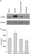
ERp46 supports in vitro tumor growth. (A) ERp46-overexpressing subclone of human PC 22Rv1 cells (ERp46+) demonstrated a 4-fold increase in ERp46 protein, while an 89% knockdown of ERp46 protein expression was observed in cells stably expressing shRNA specific for ERp46 (shERp46), compared with the corresponding control transfected cells. ERp46 was detected by western blot analysis. The expression level of β-actin served as a loading control. (B) Cell number of ERp46-manipulated subclones of 22Rv1 following 6 days of growth when seeded at 1,000 cells/well in a 96-well plate. The error bars indicate standard error of the mean. *P<0.05, compared with the corresponding control. PC, prostate cancer; sh, short hairpin; scr, scrambled; ERp46, endoplasmic reticulum protein 46.
Figure 3.
Figure 3.
ERp46 supports in vivo tumor growth. (A) Longitudinal tumor growth from mice (n=10/group) subcutaneously injected with shERp46-22Rv1 cells (white squares) was significantly reduced compared with mice injected with cells stably transfected with scrambled control shRNA (black triangles; P<0.0005). The error bars indicate standard error of the mean. (B) Tumor volume from mice (n=10/group) injected with ERp46+-22Rv1 cells (black diamonds) was significantly increased compared with mice injected with parental 22Rv1 cells (white circles; P=0.02). The error bars indicate standard error of the mean. (C) Serum PSA values from mice subcutaneously injected with shERp46-22Rv1 cells were significantly reduced compared with mice injected with cells stably transfected with scrambled control shRNA (P=0.0002). Individual values are represented by white circles and the mean by black diamonds; the box indicates the interquartile range. (D) Microvessels as demonstrated by immunohistochemistry of scrambled control 22Rv1 cell xenografts for CD31. Magnification, ×400. (E) Immunohistochemistry of shERp46 xenografts for CD31. Magnification, ×400. (F) Serum PSA values from mice subcutaneously injected with ERp46-overexpressing 22Rv1 cells compared with parental 22Rv1 cells. Individual values are represented by white circles and the mean by black diamonds; the box indicates the interquartile range. sh, short hairpin; PSA, prostate-specific antigen; CD, cluster of differentiation; ERp46, endoplasmic reticulum protein 46.
Figure 4.
Figure 4.
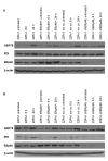
Treatment of shERp46-expressing cells with tunicamycin does not lead to an increase in PDI. (A) ERp46, GRP78 and PDI protein expression levels in ERp46-manipulated 22Rv1 cells following thapsigargin (60 nM) treatment for 6 and 24 h as determined by western blot analysis. (B) ERp46, GRP78 and PDI protein expression levels in ERp46-manipulated 22Rv1 cells following tunicamycin (2.5 µg/ml) treatment for 6 and 24 h. The expression levels of β-actin served as the loading control. sh, short hairpin; PDI, protein disulfide isomerase; scr co, scrambled control; ERp46, endoplasmic reticulum protein 46.

References

    1. Hatahet F, Ruddock LW. Protein disulfide isomerase: A critical evaluation of its function in disulfide bond formation. Antioxid Redox Signal. 2009;11:2807–2850. doi: 10.1089/ars.2009.2466. - DOI - PubMed
    1. Duivenvoorden WC, Paschos A, Hopmans SN, Austin RC, Pinthus JH. Endoplasmic reticulum protein ERp46 in renal cell carcinoma. PLoS One. 2014;9:e90389. doi: 10.1371/journal.pone.0090389. - DOI - PMC - PubMed
    1. Benham AM. The protein disulfide isomerase family: Key players in health and disease. Antioxid Redox Signal. 2012;16:781–789. doi: 10.1089/ars.2011.4439. - DOI - PubMed
    1. Charlton HK, Webster J, Kruger S, Simpson F, Richards AA, Whitehead JP. ERp46 binds to AdipoR1, but not AdipoR2, and modulates adiponectin signalling. Biochem Biophys Res Commun. 2010;392:234–239. doi: 10.1016/j.bbrc.2010.01.029. - DOI - PubMed
    1. Jessop CE, Watkins RH, Simmons JJ, Tasab M, Bulleid NJ. Protein disulphide isomerase family members show distinct substrate specificity: P5 is targeted to BiP client proteins. J Cell Sci. 2009;122:4287–4295. doi: 10.1242/jcs.059154. - DOI - PMC - PubMed