

Combination of celecoxib and PD184161 exerts synergistic inhibitory effects on gallbladder cancer cell proliferation
- PMID: 28521485
- PMCID: PMC5431146
- DOI: 10.3892/ol.2017.5914
Combination of celecoxib and PD184161 exerts synergistic inhibitory effects on gallbladder cancer cell proliferation
Abstract
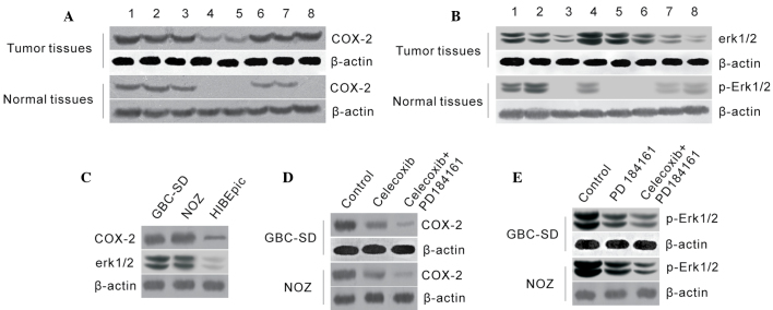
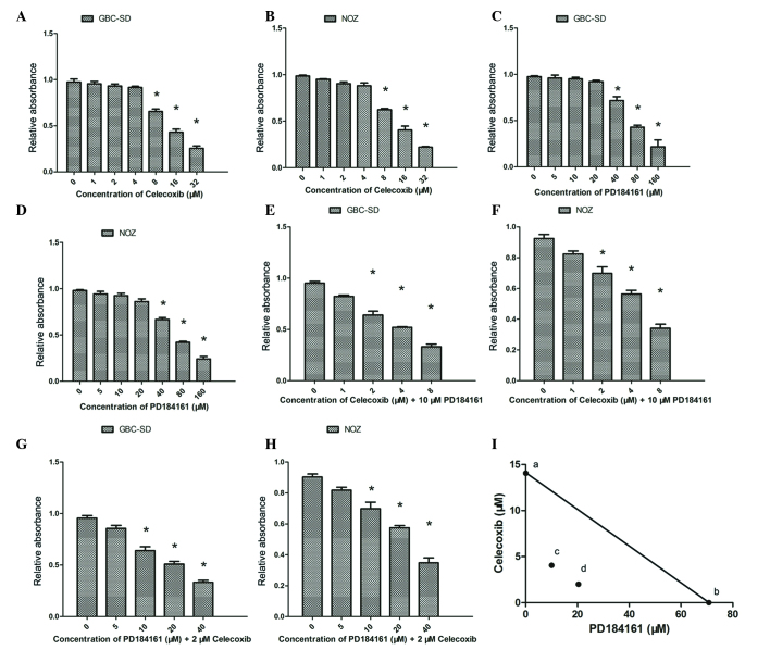
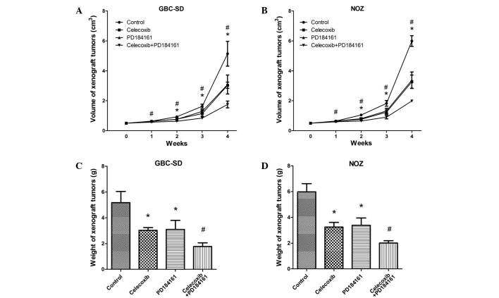
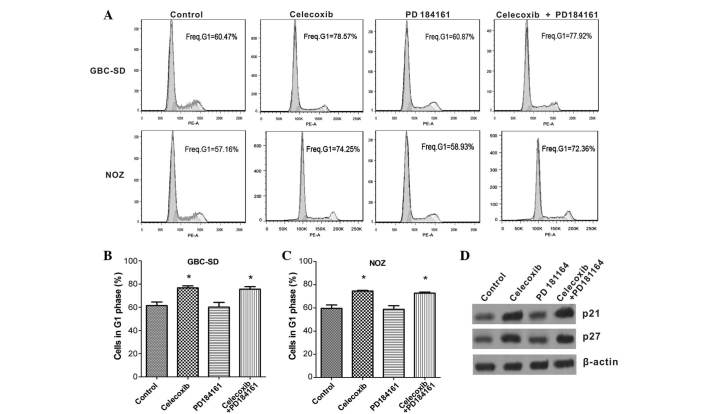
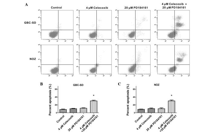
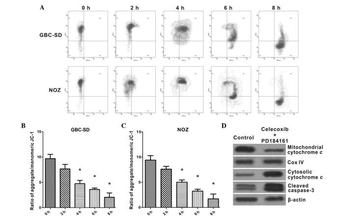
Cyclooxygenase-2 (COX-2) and extracellular signal-regulated kinase 1/2 (ERK1/2) may serve as potential targets in various types of cancer; however, the roles of these proteins in gallbladder carcinoma (GBC) have not been reported previously. In the present study, the expression levels of COX-2 and phospho (p)-ERK1/2 in GBC were examined and the biological activities of celecoxib and PD184161 (specific inhibitors of COX-2 and p-ERK1/2, respectively) on the proliferation, cell cycle and apoptosis of the GBC-SD and NOZ human GBC cell lines were evaluated by a series of in vitro and in vivo studies. COX-2 and p-ERK1/2 protein expression levels were found to be significantly elevated in GBC tissues as well as in GBC-SD and NOZ cells. Treatments with celecoxib and PD184161 significantly inhibited GBC-SD and NOZ cell growth in a concentration-dependent manner, and their combination produced a synergistic inhibitory effect. In addition, celecoxib and PD184161 significantly inhibited tumor growth in xenograft nude mice. Celecoxib treatment led to G1 arrest via the upregulation of p21 and p27 expression in GBC-SD and NOZ cells, whereas PD184161 did not affect cell cycle distribution. The combination of celecoxib and PD184161 was able to promote cell apoptosis by triggering a collapse of mitochondrial membrane potential and activating caspase-3-mediated apoptosis. In conclusion, COX-2 and p-ERK1/2 protein may serve as potential targets for GBC chemotherapy, and the combination of celecoxib and PD184161 could significantly inhibit GBC cell growth, induce cell G1 arrest and trigger cell apoptosis of GBC cells.
Keywords: PD184161; apoptosis; celecoxib; gallbladder carcinoma.
Figures
Similar articles
-
[Artemisinin inhibits proliferation of gallbladder cancer cell lines through triggering cell cycle arrest and apoptosis].Zhonghua Wai Ke Za Zhi. 2016 Mar 1;54(3):222-7. doi: 10.3760/cma.j.issn.0529-5815.2016.03.014. Zhonghua Wai Ke Za Zhi. 2016. PMID: 26932893 Chinese.
-
Inhibitory effects of deleted in liver cancer 1 gene on gallbladder cancer growth through induction of cell cycle arrest and apoptosis.J Gastroenterol Hepatol. 2014 May;29(5):964-72. doi: 10.1111/jgh.12486. J Gastroenterol Hepatol. 2014. PMID: 24329682
-
Inhibitory effect of norcantharidin on the growth of human gallbladder carcinoma GBC-SD cells in vitro.Hepatobiliary Pancreat Dis Int. 2007 Feb;6(1):72-80. Hepatobiliary Pancreat Dis Int. 2007. PMID: 17287171
-
UHRF1 depletion suppresses growth of gallbladder cancer cells through induction of apoptosis and cell cycle arrest.Oncol Rep. 2014 Jun;31(6):2635-43. doi: 10.3892/or.2014.3145. Epub 2014 Apr 23. Oncol Rep. 2014. PMID: 24756644
-
Vascular endothelial growth factor-D promotes growth, lymphangiogenesis and lymphatic metastasis in gallbladder cancer.Cancer Lett. 2012 Jan 28;314(2):127-36. doi: 10.1016/j.canlet.2011.09.004. Epub 2011 Sep 22. Cancer Lett. 2012. PMID: 22071224
Cited by
-
Arachidonic acid metabolism as a novel pathogenic factor in gastrointestinal cancers.Mol Cell Biochem. 2025 Feb;480(2):1225-1239. doi: 10.1007/s11010-024-05057-2. Epub 2024 Jul 4. Mol Cell Biochem. 2025. PMID: 38963615 Review.
-
Associations between serum uric acid and hepatobiliary-pancreatic cancer: A cohort study.World J Gastroenterol. 2020 Nov 28;26(44):7061-7075. doi: 10.3748/wjg.v26.i44.7061. World J Gastroenterol. 2020. PMID: 33311950 Free PMC article.
-
Non-Steroidal Anti-Inflammatory Drugs Increase Cisplatin, Paclitaxel, and Doxorubicin Efficacy against Human Cervix Cancer Cells.Pharmaceuticals (Basel). 2020 Dec 15;13(12):463. doi: 10.3390/ph13120463. Pharmaceuticals (Basel). 2020. PMID: 33333716 Free PMC article.
-
Gallbladder cancer: review of a rare orphan gastrointestinal cancer with a focus on populations of New Mexico.BMC Cancer. 2018 Jun 18;18(1):665. doi: 10.1186/s12885-018-4575-3. BMC Cancer. 2018. PMID: 29914418 Free PMC article. Review.
References
-
- Bonet B, eltrán M, Allal AS, Gich I, Solé JM, Carrió I. Is adjuvant radiotherapy needed after curative resection of extrahepatic biliary tract cancers? A systematic review with a meta-analysis of observational studies. Cancer Treat Rev. 2012;38:111–119. doi: 10.1016/j.ctrv.2011.05.003. - DOI - PubMed
LinkOut - more resources
Full Text Sources
Other Literature Sources
Research Materials
Miscellaneous